22.5 PUTTING IT ALL TOGETHER: GENE REGULATION IN DEVELOPMENT

The patterns of gene regulation that bring about development from a zygote to a multicellular animal or plant are complex and intricately coordinated. Development requires transitions in protein composition and in morphology that depend on tightly coordinated changes in expression of the genome.

How is a complex organism produced, with its many tissues and organs and appendages, from a single cell? Some clues can be found in that single cell—the fertilized egg. More genes are expressed during early development than at any other stage of the life cycle. For example, in the sea urchin, an oocyte (an immature egg cell) has about 18,500 different mRNAs, compared with about 6,000 different mRNAs in the cells of a typical differentiated tissue. The mRNAs in the oocyte give rise to a cascade of events that regulate the expression of many genes across both space and time.

The regulatory mechanisms used in development encompass all of the regulatory processes discussed in Chapter 21 and in this chapter thus far. Transcriptional regulation occurs, but posttranscriptional regulatory processes are particularly important.

Development Depends on Asymmetric Cell Divisions and Cell-Cell Signaling

If all cells divided to produce two identical daughter cells, multicellular organisms could never be more than a ball of identical cells. Programmed asymmetric cell divisions are required for different cell fates. Cell-cell signaling also helps guide the eventual differentiation of tissues and organs with various functions. Asymmetry in the developing embryo is thus created in several ways (Figure 22-21).

Figure 22-21: Several ways to generate asymmetry in a developing embryo. (a) Intrinsic asymmetry reflects the existing distribution of cellular components, especially mRNA and protein. The asymmetries are either inherent to the developing oocyte or created during fertilization. (b) Extrinsic asymmetry is generated by cell-cell signaling. Although asymmetry is not necessarily created in any given cell, the cell-cell signals alter the fate of a cell or a group of cells in the embryo, contributing to embryonic asymmetry. The signals can involve direct cell-cell contacts or the action of a secreted, diffusible signal.

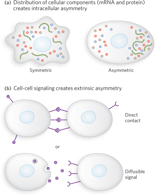
Asymmetry within the cells themselves takes the form of gradients of mRNAs and proteins that define critical axes (posterior-anterior, dorsal-ventral). In the developing oocyte, some gradients are established by deposition of mRNAs at one end or the other. Active transport in the cell can also contribute to generating a gradient. Fertilization can trigger events that create additional gradients in the fertilized egg (zygote). In many organisms, these gradients dictate different cell fates even for the daughter cells of the first cell division.

It is not enough to create a gradient in the cell, however. The mitotic spindle must also be aligned along the same axis as the gradient, so that the cell division occurs on an axis perpendicular to the gradient (see Chapter 2 for a reminder about mitotic cell division). Ensuring the proper alignment of the mitotic spindle in particular cell divisions is the function of some proteins critical to development.

In the developing embryo, cell-cell signaling generates additional asymmetry as development proceeds (see Figure 22-21). Direct contact between the lipids and glycoproteins on one cell surface and the receptors on another can guide changes in gene expression in the receptor-bearing cell. Some signals act at longer distances: diffusible molecules secreted by one cell or group of cells and detected by receptors on another, distant cell or group of cells. Ever more complex networks of signaling molecules and gene regulators are created as development proceeds.

782

Characteristic Stages of Development Several organisms became important model systems for the study of development because they are easy to maintain in a laboratory and have relatively short generation times. These include nematodes, fruit flies, zebra fish, mice, and the plant Arabidopsis thaliana (see the Model Organisms Appendix). The discussion here focuses on developmental pathways in the fruit fly. Our understanding of the molecular events during development of Drosophila melanogaster is especially well advanced and can be used to illustrate patterns and principles of general significance, and highlight the mechanisms of gene regulation that govern this complex process.

Multicellular eukaryotes develop in a process that begins with the union of an egg and a sperm cell by fertilization, to create a zygote. The egg cell has been preprogrammed by the deposition of maternal mRNAs in gradients, such that concentrations of certain maternal mRNAs vary greatly from one end of the oocyte to the other. On fertilization, cell division begins. Early in development, the fate of particular cells is determined by the concentration of maternal mRNAs, as well as by the actions of regulatory genes. As development proceeds, cascades of regulatory genes guide the various cell lineages as different tissue types develop. Although the regulatory genes are numerous, they generally fall into a small number of classes that are highly conserved, from nematodes to fruit flies to humans. Signaling pathways and processes are also highly conserved.

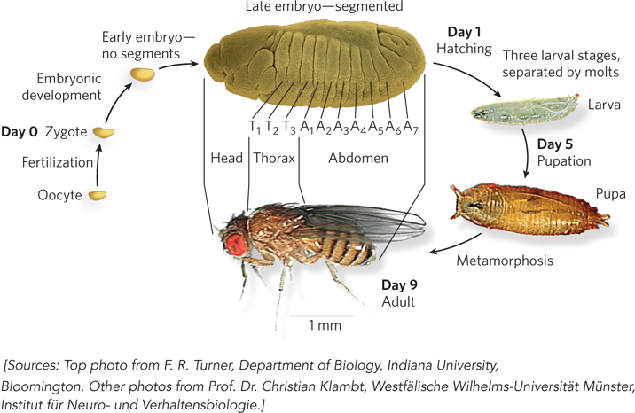
The life cycle of the fruit fly is relatively complex, and the patterns are conserved in a wide range of multicellular eukaryotes. Complete metamorphosis occurs during progression from embryo to adult fly (Figure 22-22). The final structure of the adult is forecast by features that are evident in the embryo at a very early stage. One of the most important characteristics of the embryo is its polarity: the anterior and posterior ends of the animal are readily distinguished, as are its dorsal and ventral surfaces. The fly embryo also exhibits the key characteristic of metamerism, division of the body into serially repeating segments, each with characteristic features. During development, these segments become organized into head, thorax, and abdomen. Each segment of the adult thorax has a different set of appendages. The development of this complex pattern is genetically controlled, and pattern-regulating genes—almost all with close homologs, from nematodes to humans—dramatically affect the organization of the body.

Figure 22-22: The fruit fly life cycle. The adult Drosophila is radically different in form from its immature stages, a transformation that requires extensive alterations during development. By the late embryonic stage, segments have formed, each containing specialized structures from which the various appendages and other features of the adult fly will develop. The segmented late embryo is enlarged compared with the other stages, to show detail.

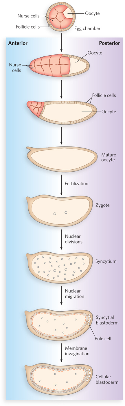
The Drosophila egg, with its 15 nurse cells, is surrounded by a layer of follicle cells (Figure 22-23). As the oocyte matures (before fertilization), mRNAs and proteins originating in the nurse and follicle cells are deposited in the egg cell, where many play a crucial role in development. After the fertilized egg is laid, the nucleus divides and the nuclear descendants continue to divide in synchrony every 6 to 10 minutes. Plasma membranes are not formed around the nuclei, which are distributed within the egg cytoplasm, forming a syncytium. During rounds 8 to 11 of nuclear division, the nuclei migrate to the egg’s outer layer, forming a monolayer surrounding the common yolk-rich cytoplasm; this is the syncytial blastoderm. After a few additional divisions (producing up to 6,000 nuclei), membrane infoldings create a layer of cells, forming the cellular blastoderm. At this stage, the mitotic cycles in the cells lose their synchrony. The developmental fate of the cells is determined by the mRNAs and proteins originally deposited in the egg by the nurse and follicle cells.

Figure 22-23: Early development in Drosophila. During oocyte development, maternal mRNAs and proteins are deposited in the oocyte by nurse cells and follicle cells. After fertilization, nuclear divisions occur in synchrony in the common cytoplasm (syncytium), and nuclei migrate to the periphery. Membrane invaginations surround the nuclei to create a monolayer of cells at the periphery; this is the cellular blastoderm stage. During the early nuclear divisions, several nuclei at the far posterior of the embryo become pole cells, which later become the germ-line cells.

783

Cascades of Regulatory Proteins in Development The role of key genes in development is to regulate other genes. Temporal and spatial regulation is critical to the gradual maturation of cells and tissues as cell divisions continue from embryo to adult. As each successive layer of regulatory genes is activated, the embryo acquires a finer specialization of cellular function.

Several types of RNAs and proteins in the early embryo, and proteins with essential roles in later stages of development, follow patterns widely conserved in multicellular eukaryotes. As defined by Christiane Nüsslein-Volhard, Edward B. Lewis, and Eric F. Wieschaus for Drosophila, three major classes of pattern-regulating genes—maternal, segmentation, and homeotic genes—function in successive developmental stages to specify the basic features of the fruit fly embryo body.

Maternal genes are expressed in the unfertilized egg, and the resulting maternal mRNAs remain dormant until fertilization. Maternal mRNAs provide most of the required proteins in the very early stages of development, and in fruit flies, this occurs until the cellular blastoderm forms. Some of the proteins encoded by maternal mRNAs direct the spatial organization of the developing embryo to establish its polarity. Segmentation genes, transcribed after fertilization, direct the formation of the proper number of body segments. In nematodes, similar genes guide the formation of specific tissues following completion of the earliest stages of embryogenesis. At least three subclasses of segmentation genes act at successive stages of Drosophila development. Gap genes divide the developing embryo into several broad regions, and pair-rule genes, along with segment polarity genes, define 14 stripes that become the 14 segments of a normal fly embryo. Homeotic genes, expressed at a later stage, specify the organs and appendages that will develop in particular body segments.

784

The many regulatory genes in these three classes direct the development of an adult organism, with a head, thorax, and abdomen, the proper number of segments, and the correct appendages on each segment. Although fruit fly embryogenesis takes about a day to complete, all these genes are activated during the first 4 hours. During this period, some mRNAs and proteins are present for only a few minutes at specific points in time. Some of the genes code for transcription factors that affect the expression of other genes in a kind of developmental cascade. Regulation at the level of translation also occurs, and many of the regulatory genes encode translational repressors, most of which bind to the 3′UTR of mRNAs. Because many mRNAs are deposited in the egg long before their translation is required, translational repression is especially important for regulation in developmental pathways.

Early Development Is Mediated by Maternal Genes

In invertebrates, a prescribed developmental path is evident from the very first embryonic cell division. The nonequivalence of the daughter cells of this first division implies a structural and functional asymmetry in the fertilized egg. The asymmetry is mediated by established gradients of molecules called morphogens—mRNAs and proteins produced by maternal genes.

In Drosophila, some maternal genes are expressed within the nurse and follicle cells, and some in the egg itself. In the unfertilized egg, the maternal gene products establish the critical anterior-posterior and dorsal-ventral axes, thereby defining which regions of the radially symmetric egg will develop into the head and abdomen and the top and bottom of the adult fly. A key event in very early development is establishing mRNA and protein gradients along the body axes. Some maternal mRNAs have protein products that diffuse through the cytoplasm, creating an asymmetric distribution in the egg. Various cells in the cellular blastoderm therefore inherit different amounts of these proteins, setting the cells on different developmental paths. The products of the maternal mRNAs include transcription activators or repressors as well as translational repressors, all regulating the expression of other pattern-regulating genes. Thus, the resulting gene expression sequences and patterns differ among cell lineages, ultimately orchestrating the development of each adult structure.

The anterior-posterior axis in Drosophila is also partially defined by the transcription factors produced by the bicoid and nanos genes. The bicoid mRNA is synthesized by nurse cells and deposited in the unfertilized egg near its anterior pole. Nüsslein-Volhard found that this mRNA is translated soon after fertilization, and the Bicoid protein diffuses through the cell to create, by the seventh nuclear division, a concentration gradient radiating out from the anterior pole (Figure 22-24a).

Figure 22-24: Distribution of a maternal gene product in a Drosophila egg. (a) The micrograph of an immunologically stained egg shows the distribution of the bicoid (bcd) gene product, the Bicoid protein. The graph shows stain intensity (protein concentration) along the length of the egg. This distribution is essential for normal development of the anterior structures. (b) If the bcd gene is not expressed by the mother (a bcd/bcd mutant), no bicoid mRNA is deposited in the egg, resulting in lack of Bicoid protein, as seen in the micrograph. The resulting embryo has two posteriors (and soon dies).

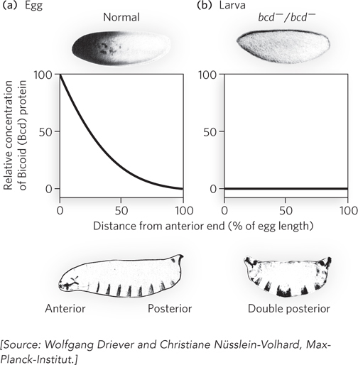
Bicoid contains a homeodomain (see Chapter 19). As a transcription activator, Bicoid activates the expression of several segmentation genes. It is also a translational repressor that inactivates certain mRNAs. The amounts of Bicoid in various parts of the embryo affect the subsequent expression of other genes in a threshold-dependent way. Genes are transcriptionally activated or translationally repressed only where the concentration of Bicoid exceeds the threshold. Bicoid plays a critical role in anterior development. The absence of Bicoid results in development of an embryo with two abdomens but no head and no thorax (Figure 22-24b). Embryos without Bicoid can develop normally if an adequate amount of bicoid mRNA is injected into the egg at the appropriate end.

The nanos gene has an analogous role, but its mRNA is deposited at the posterior end of the egg, and the anterior-posterior Nanos protein gradient peaks at the posterior pole. Nanos is a translational repressor, conserved from worms to humans.

785

A broader view of the effects of maternal genes in Drosophila reveals a more precise picture of a developmental circuit. In addition to bicoid and nanos mRNAs, deposited in the egg asymmetrically, several other maternal mRNAs are deposited uniformly throughout the egg cytoplasm. Three of them encode the Pumilio, Hunchback, and Caudal proteins—all affected by Nanos and Bicoid (Figure 22-25).

Figure 22-25: Regulatory circuits of the anterior-posterior axis in a Drosophila egg. The bicoid and nanos mRNAs are localized near the anterior and posterior poles of the egg, respectively. The caudal, hunchback, and pumilio mRNAs are distributed throughout the cytoplasm. Gradients of Bicoid and Nanos proteins lead to accumulation of Hunchback protein in the egg’s anterior region and Caudal protein in its posterior. Because Pumilio requires Nanos for its activity as a translational repressor of hunchback mRNA, Pumilio functions only at the posterior end.

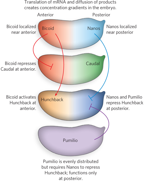
Caudal and Pumilio are involved in the development of the fruit fly’s posterior end. Caudal is a transcription activator with a homeodomain; Pumilio is a translational repressor from the PUF family of proteins (see Figure 22-28). Hunchback plays an important part in developing the anterior end; it is also a transcription factor for several genes, in some cases an activator and in others a repressor. Bicoid suppresses the translation of caudal mRNA at the anterior end and also acts as a transcription activator of hunchback mRNA in the cellular blastoderm. Because hunchback is expressed through maternal mRNAs and from genes in the developing egg, it is considered a maternal as well as a segmentation gene. The result of Bicoid’s activities is an increased concentration of Hunchback at the anterior end of the egg. Nanos and Pumilio act as translational repressors of hunchback, suppressing synthesis of Hunchback near the posterior end of the egg. Pumilio does not function in the absence of Nanos, and the gradient of nanos expression confines the activity of both proteins to the posterior region. Translational repression of the hunchback gene leads to degradation of hunchback mRNA near the posterior end. However, a lack of Bicoid in the posterior leads to expression of caudal. In this way, the Hunchback and Caudal proteins become asymmetrically distributed in the egg.

Segmentation Genes Specify the Development of Body Segments and Tissues

Segmentation genes are the zygotic genes that take over after maternal genes. Many operate at the level of transcriptional regulation. Gap genes, pair-rule genes, and segment polarity genes are activated in a cascadelike sequence at successive stages of embryonic development. The expression of gap genes is generally regulated by the products of one or more maternal genes. Gap genes activate the pair-rule genes, which in turn activate the segment polarity genes. This cascade of gene expression is accompanied by the gradual formation of 14 parasegments, then the true segments. Only a few cells (or nuclei) wide, parasegments are delimited by temporary grooves. Segments are offset from parasegments, so that each segment later encompasses the anterior part of a parasegment and the posterior part of the adjacent one. The anterior segments eventually fuse to form the head.

Pair-rule genes are expressed in alternating parasegments, and one well-characterized segmentation gene in the pair-rule subclass is fushi tarazu (ftz). When ftz is deleted, the embryo develops 7 segments instead of the normal 14, each segment twice the usual width. The Fushi-tarazu protein (Ftz) is a transcription activator with a homeodomain. The mRNAs and proteins derived from the ftz gene accumulate in a striking pattern of seven stripes that encircle the posterior two-thirds of the embryo (Figure 22-26). The stripes demarcate half of the parasegments; the development of alternating segments is compromised if ftz function is lost. The Ftz protein and a few similar regulatory proteins directly or indirectly regulate the expression of vast numbers of genes in the continuing developmental cascade.

Figure 22-26: Distribution of the fushi tarazu (ftz) and even-skipped (eve) gene products in early Drosophila embryos. (a) The ftz gene product can be detected in seven bands around the circumference of the embryo. These alternate with bands where the eve gene is expressed. (b) In a cross-sectional autoradiograph, the ftz bands appear as dark spots (generated by a radioactive label) and demarcate the anterior margins of the segments that will appear in the late embryo.

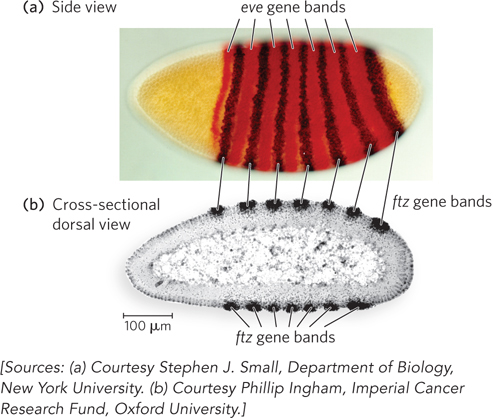
In the stripes where ftz is repressed, repression is mediated in part by another pair-rule gene called even-skipped (eve) (see Figure 21-15). The expression of eve is activated by Bicoid and Hunchback, and is highest in the parasegments where ftz gene expression is low. This gives rise to the alternating pattern of ftz and eve expression. The expression of eve is repressed by two other gap genes, krüppel and giant. The alternating pattern of eve and ftz expression is due to variations in gap gene expression from one parasegment to the next.

786

Gap and pair-rule genes operate during the first 2.5 hours of Drosophila development, when the embryo is still a syncytium. Virtually all these genes encode transcription factors, which have localized access to the nuclei in the syncytium. Segment polarity genes, the last group in the regulatory cascade, act at a stage when cells have formed. Some of the focus now shifts from transcription factors to cell-cell signaling pathways. The signaling helps reinforce the alternating boundaries between parasegments, then segments. Further, the function of adjacent segments is made interdependent by the pattern of segment polarity gene expression. A key example can be seen in the genes wingless (wg), engrailed (en), and hedgehog (hh). Wingless (Wg) and Engrailed (En) proteins are initially expressed in alternating cells, due to activation by pair-rule genes. However, the relevant pair-rule gene products recede after a few hours, and the continued expression of Wg and En becomes interdependent across opposite sides of parasegment boundaries (Figure 22-27).

Figure 22-27: Interdependent signaling loops across segment boundaries in Drosophila. Part of the maintenance of parasegment, and later segment, boundaries involves cell-cell signaling. Wnt-class signaling with the Wg protein, the wingless (wg) gene product, induces expression of the engrailed (en) gene in recipient cells. The En protein triggers expression of hedgehog, part of a non–Wnt-class signaling pathway. The Hedgehog protein (Hh) promotes expression of Wg in the original cells and completes the closed signaling loop.

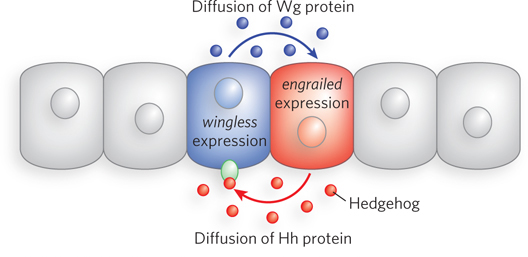
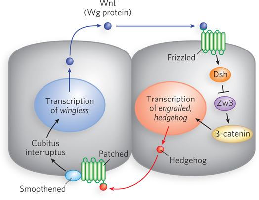
The Wg protein is a signal of the Wnt class, and studies of the wg gene helped define the Wnt-class signaling pathways, which play key roles in development in eukaryotes, from nematodes to humans (the name “Wnt,” wingless type, originated in the study of a mouse gene homologous to the Drosophila gene wingless). Wnt-class pathways generally consist of a secreted Wnt glycoprotein that constitutes the signal, one or more proteins involved in the secretion process, and a receptor protein in the membrane of the target cell (Figure 22-28). Additional proteins act as regulators.

Figure 22-28: A Wnt-class signaling pathway in Drosophila development. The Wnt signal is secreted from one cell and interacts with a receptor on another cell. The signal in this case results in gene activation, via a pathway that uses a receptor (Frizzled) and signaling proteins Dsh, Zw3, and β-catenin. The secreted Hedgehog protein also works through a signaling pathway, involving a different receptor and different set of signaling proteins, as shown. Cubitus interruptus is a zinc finger transcription factor involved in Hedgehog signaling; Smoothened is a membrane receptor for Hedgehog; and Patched is a protein that regulates the expression and function of Smoothened.

787

Wnt proteins are highly homologous from one species to the next. They generally have a nearly invariant pattern of 23 Cys residues (some of which may form disulfide bonds needed for folding), an N-terminal signal sequence that helps guide secretion, and several N-glycosylation sites. They also have at least two lipid modifications: a palmitoyl group is added at a conserved Cys residue, and a palmitoleyl group at a conserved Ser. Wnt proteins are synthesized, and the lipid modifications are made, in the endoplasmic reticulum. The proteins move through a normal secretion pathway, from ER to Golgi complex, and are then transported to the cell surface in vesicles. Some of the secreted Wnt proteins are associated with lipoprotein particles.

The Wg protein is modified with lipids by an acyltransferase in the endoplasmic reticulum that is encoded by the gene porcupine. In nearby cells on the opposite side of the adjacent parasegment boundary, the secreted Wg protein interacts with a receptor that is the product of the gene frizzled (fz). In the recipient cell, the interaction triggers a signaling pathway that ultimately results in expression of the En protein. En is a transcription activator of the hedgehog gene. The Hedgehog (Hh) protein, part of a non–Wnt-class signaling pathway, is secreted and interacts with receptors on the Wg-producing cells. The resulting signal activates more Wg protein synthesis. The entire cycle is self-sustaining and self-reinforcing. Most of the other known segment polarity genes encode proteins that are part of either the wingless (Wnt) or the hedgehog (Hh) signaling pathway. In the alternating segments, the En protein and other transcription factors activate or repress a series of additional genes that now begin to give each segment a distinctive function. Many of these targets are homeotic genes.

Homeotic Genes Control the Development of Organs and Appendages

A set of 8 to 11 homeotic genes directs the formation of structures at specific locations in the body plan of most multicellular eukaryotes. Fewer homeotic genes are present in some simple eukaryotes. Even yeast has two homologs, regulators of mating-type switching (see Chapter 21). These genes are now more commonly referred to as Hox genes (from homeobox, the conserved gene sequence that encodes the homeodomain). However, these are not the only development-related proteins to include a homeodomain (as noted above, Bicoid has a homeodomain), and Hox is more a functional than a structural classification.

Hox genes are sometimes organized in genomic clusters. Drosophila has one such cluster, and mammals have four. The order of genes within the clusters is colinear with their targets of action, from the anterior to the posterior of the developing embryo. In Drosophila, each Hox gene is expressed in a particular embryonic segment and controls the development of the corresponding part of the mature fly (Figure 22-29a). The terminology for describing Hox genes can be confusing. They have historical names in the fruit fly (e.g., ultrabithorax), whereas in mammals they are designated by two competing systems based on lettered (A, B, C, D) or numbered (1, 2, 3, 4) clusters (Figure 22-29b).
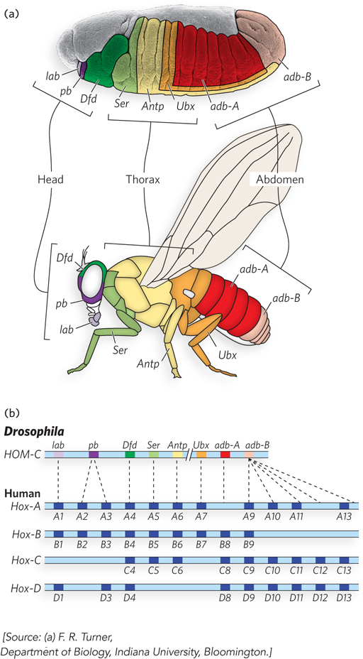
Figure 22-29: The Hox gene clusters and their effects on development. (a) Each Hox gene in the fruit fly directs the development of structures in a defined part of the body and is expressed in defined regions of the embryo (coded by color). (b) Drosophila has one Hox gene cluster (HOM-C). The genes are color-coded to match the fly segments in (a). The human genome has four Hox gene clusters (Hox-A through Hox-D). Many Hox genes are highly conserved in animals. Evolutionary relationships between genes in the Drosophila Hox gene cluster and those in the human (mammalian) Hox gene clusters are indicated by dashed lines. Similar relationships between the four sets of human Hox genes are indicated by vertical alignment.

788

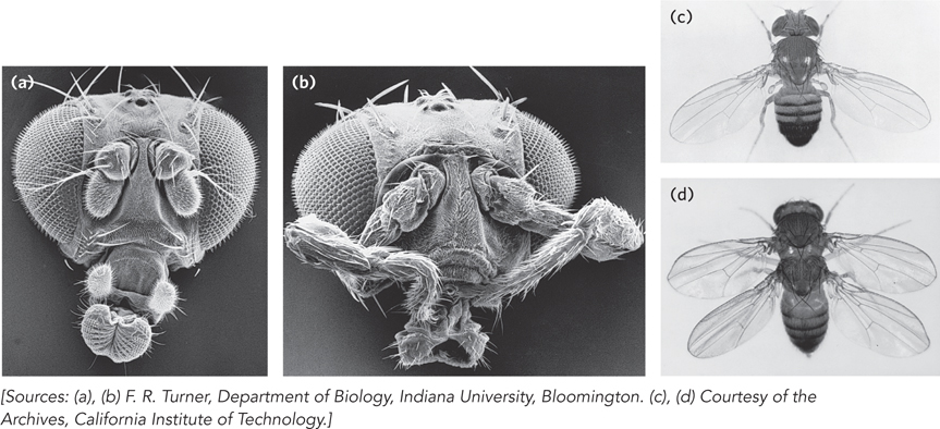
The loss of Hox genes in fruit flies, by mutation or deletion, causes the appearance of a normal appendage or body structure at an inappropriate body position. An important example is the ultrabithorax (ubx) gene. When the Ubx protein function is lost, the first abdominal segment develops incorrectly, with the structure of the third thoracic segment. Other known homeotic mutations cause the formation of an extra set of wings, or two legs at the position in the head where the antennae are normally found (Figure 22-30). The Hox genes often span long regions of DNA. The ubx gene, for example, is 77,000 bp long. More than 73,000 bp are in introns, one of which is more than 50,000 bp long. Transcription of the ubx gene takes nearly an hour. The delay this imposes on ubx gene expression is believed to be a timing mechanism involved in the temporal regulation of subsequent steps in development. Many Hox genes are further regulated by miRNAs encoded by intergenic regions of the Hox gene clusters. All Hox gene products are themselves transcription factors that regulate the expression of an array of downstream genes.

Figure 22-30: Effects of Hox gene mutations in Drosophila. (a) Normal head structure. (b) Homeotic mutant (antennapedia) in which antennae are replaced by legs. (c) Normal body structure. (d) Homeotic mutant (bithorax) in which a segment has developed incorrectly to produce an extra set of wings.

The conservation of some Hox genes is extraordinary. For example, the products of the homeobox-containing Hoxa-7 gene in mice and antennapedia gene in fruit flies differ in only one amino acid residue. Of course, although the molecular regulatory mechanisms may be similar, many of the ultimate developmental events are not conserved (humans do not have wings or antennae). The different outcomes are brought about by variance in the downstream target genes controlled by the Hox genes (see the How We Know section at the end of this chapter). The discovery of structural determinants with identifiable molecular functions is the first step in understanding the molecular events underlying development. As more genes and their protein products are discovered, the biochemical side of this vast puzzle will be elucidated in increasingly rich detail.

Stem Cells Have Developmental Potential That Can Be Controlled

If we can understand development, and the mechanisms of gene regulation behind it, we can control it. An adult human has many different types of tissues. Many of the cells are terminally differentiated and no longer divide. If an organ malfunctions due to disease, or a limb is lost in an accident, the tissues are not readily replaced. Most cells, because of the regulatory processes that are in place, or even the loss of some or all genomic DNA, are not easily reprogrammed. Medical science has made organ transplants possible, but organ donors are a limited resource, and organ rejection remains a major medical problem. If humans could regenerate their own organs or limbs or nervous tissue, rejection would no longer be an issue. Real cures for kidney failure or neurodegenerative disorders could become reality.

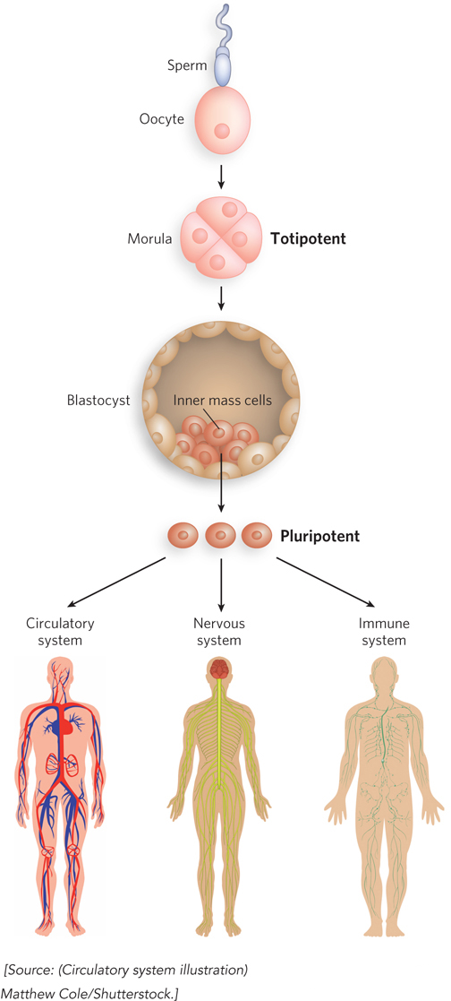
The key to tissue regeneration lies in stem cells—cells that have retained the capacity to differentiate into various tissues. In humans, after an egg is fertilized, the first few cell divisions create a ball of totipotent cells (the morula), which have the capacity to differentiate individually into any tissue or even into a complete organism (Figure 22-31). Continued cell division produces a hollow ball, a blastocyst. The outer cells of the blastocyst eventually form the placenta. The inner layers form the germ layers of the developing fetus—the ectoderm, mesoderm, and endoderm. These cells are pluripotent: they can give rise to cells of all three germ layers and can be differentiated into many types of tissues. However, they cannot differentiate into a complete organism. Some of these cells are unipotent: they can develop into only one type of cell and/or tissue. It is the pluripotent cells of the blastocyst, the embryonic stem cells, that were originally used in embryonic stem cell research.

Figure 22-31: Totipotent and pluripotent stem cells. Cells of the morula stage are totipotent and have the capacity to differentiate into a complete organism. The source of pluripotent embryonic stem cells is the inner mass cells of the blastocyst. Pluripotent cells give rise to many tissue types, but they cannot form complete organisms.

789

Stem cells have two functions: to replenish themselves and, at the same time, to provide cells that can differentiate. These tasks are accomplished in multiple ways (Figure 22-32a). All or parts of the stem cell population can, in principle, be involved in replenishment, differentiation, or both.

Figure 22-32: Stem cell proliferation versus differentiation and development. Stem cells must strike a balance between self-renewal and differentiation. (a) Some possible cell division patterns that allow for replenishment of stem cells and production of some differentiated cells. Each cell may produce one stem cell and one differentiated cell, or two differentiated cells or two stem cells in defined parts of the tissue or culture. Alternatively, a gradient of growth conditions can be established, with cell fates differing from one end of the gradient to the other. (b) Establishing a developmental niche through stem cell contact with a cell or group of cells. Molecular signals (dashed arrows) provided by niche cells (in this case, for plants, a distal tip cell) help orient the mitotic spindle for stem cell division and ensure that one daughter cell retains stem cell properties.

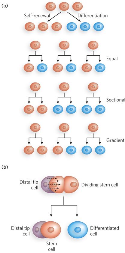
Other types of stem cells can potentially be used for medical benefit. In the adult organism, adult stem cells, as products of additional differentiation, have a more limited potential for further development than do embryonic stem cells. For example, the hematopoietic stem cells of bone marrow can give rise to many types of blood cells, as well as to cells with the capacity to regenerate bone. They are referred to as multipotent. However, these cells cannot differentiate into a liver or kidney or neuron. Adult stem cells are often said to have a niche, a microenvironment that promotes stem cell maintenance while allowing differentiation of some daughter cells as replacements for cells in the tissue they serve (Figure 22-32b). Hematopoietic stem cells in the bone marrow occupy a niche in which signaling from neighboring cells and other cues maintain the stem cell lineage. At the same time, some daughter cells differentiate to provide needed blood cells. Understanding the niche in which stem cells operate, and the signals the niche provides, is essential in efforts to harness the potential of stem cells for tissue regeneration.

790

All stem cells have problems with respect to human medical applications. Adult stem cells have a limited capacity to regenerate tissues, are generally present in small numbers, and are hard to isolate from an adult human. Embryonic stem cells have much greater differentiation potential and can be cultured to generate large numbers of cells. However, their use is accompanied by ethical concerns related to the necessary destruction of human embryos. Identifying a source of plentiful and medically useful stem cells that does not raise ethical concerns remains a major goal of medical research.

Our ability to culture stem cells (i.e., maintain them in an undifferentiated state), and to manipulate them to grow and differentiate into particular tissues, is very much a function of our understanding of developmental biology. The identification and culturing of pluripotent stem cells from human blastocysts was reported by James Thomson and his colleagues in 1998. This advance led to the long-term availability of established cell lines for research.

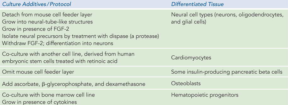
In early work, mouse and human embryonic stem cells were used for most research. Although both types of stem cells are pluripotent, they require very different culture conditions, optimized to allow cell division indefinitely without differentiation. Mouse embryonic stem cells are grown on a layer of gelatin and require the presence of leukemia inhibitory factor (LIF). Human embryonic stem cells are grown on a feeder layer of mouse embryonic fibroblasts and require basic fibroblast growth factor (bFGF or FGF-2). The use of a feeder cell layer implies that the mouse cells are providing a diffusible product or some surface signal, not yet known, that is needed by human stem cells to either promote cell division or prevent differentiation. Recent research suggests that at least one of these diffusible products may be a Wnt-class protein. Some success has been achieved in directing the differentiation of human embryonic stem cells into particular tissue types; some of the progress in stimulating stem cell differentiation is summarized in Table 22-1. However, due to limited availability and ethical concerns, as noted above, embryonic stem cells have not been an ideal system on which to base continued research and potential medical applications.

The answer is to find another, more abundant and noncontroversial source of pluripotent stem cells. In 2007, researchers first reported success in reprogramming somatic cells to pluripotency. Skin cells—first from mice, then from humans—were reprogrammed to take on the characteristics of pluripotent stem cells. The reprogramming involves manipulations to get the cells to express some or all of four transcription factors involved in development: Oct4, Sox2, Krüppel-like factor 4 (KLF4), and cMyc—collectively known as OSKM factors. All of these are known to help maintain the stem cell–like state. The result is cells called induced pluripotent stem cells, or iPS cells. These, in turn, have been used to generate a range of tissue types. Gradual improvements in this technology are continuing. A new branch of medicine is slowly emerging, called regenerative medicine, which may eventually provide new approaches to the repair of damaged tissue after heart attacks and strokes and other traumas. The use of reprogrammed stem cells derived from the same patient may eliminate tissue rejection and provide a source of new, healthy tissue.

Figure 22-1: Requirements for Differentiating Human Embryonic Stem Cells into Various Tissue Types

791

SECTION 22.5 SUMMARY

  • Development of a multicellular organism presents the most complex regulatory challenge.

  • The fate of cells in the early embryo is determined in part by establishment of anterior-posterior and dorsal-ventral gradients of proteins that act as transcription activators or translational repressors, regulating the genes required for the development of structures appropriate to a particular part of the organism.

  • Sets of regulatory genes operate in temporal and spatial succession, transforming given areas of an egg cell into predictable structures in the adult organism.

  • The developmental fate of cell lineages during development is also shaped by cell-cell signaling pathways in which signals from one cell lineage affect the fate of others. The Wnt-class signaling pathway is one well-studied example.

  • In vertebrates, stem cells retain significant developmental potential. The differentiation of stem cells into functional tissues can be controlled by extracellular signals and conditions.