2.1 The Neuron
Why are psychologists interested in how neurons work? Isn’t this biology and not psychology? The answer is that it’s both. Humans are biological organisms. To understand our behavior and mental processes, we need to understand their biological underpinnings, starting with the cellular level, the neuron. How we feel, learn, remember, and think all stem from neuronal activity. So, how a neuron works and how neurons communicate are crucial pieces of information in solving the puzzle of human behavior and mental processing.
In explaining how a neuron works, we will talk about how neurons communicate with one another. We have a fairly good understanding of how information is transmitted, but we do not have as good an understanding of exactly how these vast communication networks of neurons oversee what we do and make us what we are. These more complex questions are the remaining key pieces of the puzzle to be solved. In this section, we will cover the part of the story that is best understood—
The Structure of a Neuron
neurons Cells that transmit information within the nervous system.
glial cells (glia) Cells in the nervous system that comprise the support system for the neurons.
The brain and the nervous system are composed of two types of cells—
Recent research is also questioning the idea that glial cells merely provide a support system for neurons (Barres, 2008; Fields, 2009, 2011; Koob, 2009). It appears that not only do neurons and glial cells communicate but also that glial cells communicate with one another in a separate but parallel network to the neuronal network, influencing the brain’s performance. Glial cells also appear to influence the formation of neuronal connections and to aid in determining which neuronal connections get stronger or weaker over time, both essential to learning and to storing memories. In addition, glial cells may play an important role in mental disorders such as schizophrenia and depression and in neurodegenerative diseases such as Parkinson’s and Alzheimer’s. Whereas neuroscientists are excited by all of these possibilities and the prospect of doing research on these cells that have been largely ignored until recently, neurons are still viewed as the most important cells for communication within the human nervous system and thus will be the focus of our discussion.
Neurons all have the same basic parts and structure, and they all operate the same way. A generic neuron with all of the important parts identified is depicted in Figure 2.1. The three main components of a neuron are the dendrites, cell body, and axon. Let’s first get a general idea of the main functions of these three parts and their relationships to one another.
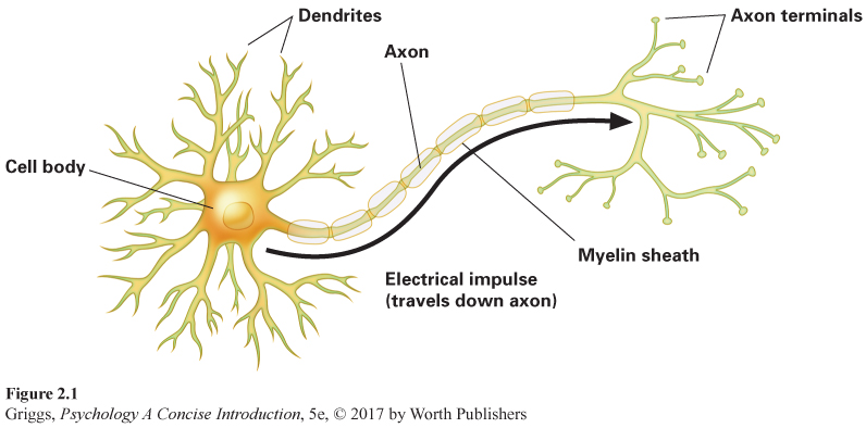
dendrites Fibers projecting out of the cell body of a neuron whose function is to receive information from other neurons.
cell body The part of the neuron that contains its nucleus and the other biological machinery to keep the cell alive and that decides whether to generate a neural impulse in order to pass incoming information on to other neurons.
axon The long, singular fiber projecting out of the cell body of a neuron, whose function is to conduct the neural impulse from the cell body to the axon terminals, triggering chemical communication with other neurons.
Dendrites are the fibers that project out of the cell body like the branches of a tree. Their main function is to receive information from other neurons. The dendrites pass this information on to the cell body, which contains the nucleus of the cell and the other biological machinery that keeps the cell alive. The cell body also decides whether to pass the information from the dendrites on to other neurons. If the cell body decides to pass along the information, it does so by way of the axon—the long, singular fiber leaving the cell body. At its end, the axon subdivides into axon terminals, branchlike endings. The main function of the axon is to conduct information from the cell body to the axon terminals in order to trigger the transmission of information with other neurons. Axons vary greatly in length, with the longest ones going from the spinal cord to the toes. Given this general understanding of the parts of the neuron, let’s look more closely at exactly how they are involved in transmitting information.
How Neurons Communicate
The first point to note in learning about how neurons communicate with one another (and sometimes with muscles and glands) is that the process is partly electrical and partly chemical. Within a neuron, it’s electrical—
The electrical impulse. The electrical part of the story begins with the messages received by the dendrites from other neurons. These inputs are either excitatory (telling the neuron to generate an electrical impulse) or inhibitory (telling the neuron not to generate an electrical impulse). The cell body decides whether to generate an impulse by continually calculating this input. If the excitatory input outweighs the inhibitory input by a sufficient amount, then the cell body will generate an impulse. The impulse travels from the cell body down the axon to the axon terminals. This impulse is an all-
myelin sheath An insulating layer covering an axon that allows for faster neural impulses.
The impulses in different neurons travel down the axon at varying rates up to around 200 miles per hour (Dowling, 1998). This may seem fast, but it is much slower than the speed of electricity or computer processing. A major factor determining the impulse speed for a particular neuron is whether its axon is encased in a myelin sheath—an insulating layer of a white fatty substance. The myelin sheath is composed of glial cells that wrap around the neuron’s axon. With no myelin sheath, the impulse travels slowly down the axon in a continuous fashion, like a fuse burning down on a stick of dynamite. The rate is faster in axons encased in myelin because the impulse can only be regenerated at the periodic gaps in the sheath where there is no myelin. A myelinated axon (like the one in Figure 2.1) looks like a string of sausages with gaps between the sausages. The impulse “leaps” from gap to gap instead of traveling continuously down the axon. To understand why this is faster, think about walking across a room in a heel-
Damage to this myelin sheath will result in serious problems. For example, multiple sclerosis causes deterioration of the myelin sheath that encases neuronal axons. This means that impulses can no longer leap down the axon, or eventually even travel down the axon, so information transmission is greatly slowed. People with multiple sclerosis experience incapacitating behavioral changes, such as difficulty in moving. Sadly, there is presently no cure for multiple sclerosis, but recent research using stem cell (an unspecialized cell in the body that can develop into a specialized cell) transplantation has found some preliminary encouraging results (Burt et al., 2015).
Given its white color, myelin is also responsible for the distinction between white matter and gray matter in the brain. Myelinated axons make up the “white” matter; unmyelinated axons, cell bodies, and dendrites make up the “gray” matter. Why the distinction? Myelination creates a whitish appearance because of the white color of the myelin. Unmyelinated parts of a neuron appear grayish. If we were able to look at the two cerebral hemispheres of the brain, they would appear grayish because we are mainly looking at the billions of cell bodies that make up their outside layer. Hence, the expression, “Use your gray matter.”
neurotransmitter A naturally occurring chemical in the nervous system that specializes in transmitting information between neurons.
synaptic gap (synapse) The microscopic gap between neurons across which neurotransmitters travel to carry their messages to other neurons.
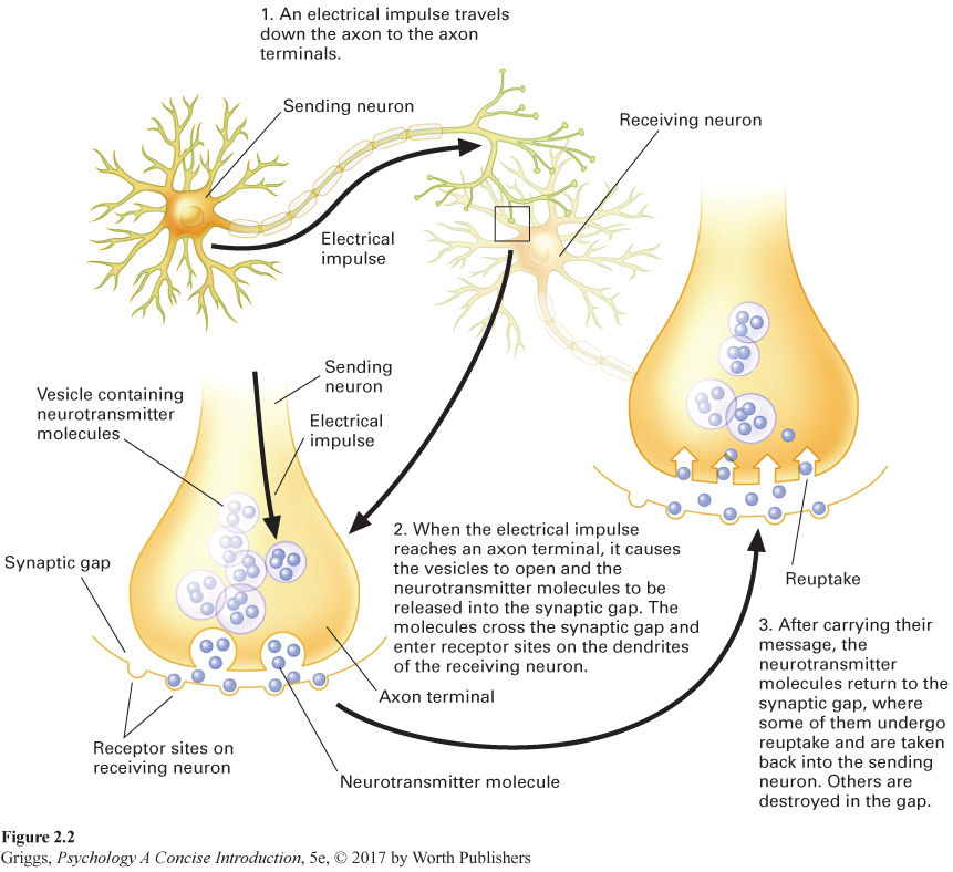
Chemical communication between neurons. What happens when the electrical impulse reaches the axon terminals? The answer is depicted in Figure 2.2. In the axon terminals, there are tiny vesicles (sacs) containing a neurotransmitter, a naturally occurring chemical in our nervous system that specializes in transmitting information. When the impulse reaches the sending neuron’s axon terminals, it causes the vesicles to open and the neurotransmitter molecules to come out and go into the synaptic gap (or synapse), the microscopic gap (less than a millionth of an inch wide) between neurons. Two thousand of these tiny synaptic gaps would fit comfortably inside the thinnest of human hairs (Lynch & Granger, 2008). The neurotransmitter molecules cross the gap and enter receptor sites on the dendrites of other neurons. This is achieved by what is termed binding—
So who discovered the synapse, which is so critical to our existence? Synapses were first documented by Spanish anatomist Santiago Ramón y Cajal in the nineteenth century (Rapport, 2005). Ramón y Cajal, often referred to as the father of neuroscience, shared the Nobel Prize in Physiology or Medicine in 1906 for his contributions to our understanding of the nervous system. However, Ramón y Cajal did not use the word synapse to refer to the microscopic gap between neurons. It was Sir Charles Scott Sherrington, another Nobel Prize winner, who coined this term. According to Rapport (2005), when Sherrington was writing material for a new edition of Michael Foster’s A Textbook of Physiology, he grew tired of having to continually invent ways to describe the conduction of an impulse across a gap to dendrites. To eliminate this problem, Sherrington coined the term synapse, which appeared for the first time in Foster’s seventh edition in 1897.
Brain scans. In order to carry out their essential communication work, neurons require oxygen and other nutrients such as blood sugars. This is why about 20% of the body’s blood supply is pumped to the brain (Gazzaniga, Ivry, & Mangun, 2002), and the brain consumes about a quarter of the body’s oxygen (Ackerman, 2004). Without oxygen, neurons die within minutes. Neurons doing more work require even more oxygen and nutrients. This fact is the key to how various types of brain scans work. There are several types of brain scans, but we will discuss two that are often used by psychology researchers.
positron emission tomography (PET) scans A visual display of the activity levels in various areas in the brain generated by detecting the amount of positron emission created by the metabolization of radioactive glucose in each area.
In positron emission tomography (PET) scans, a harmless dose of radioactive glucose (sugar) is introduced into the bloodstream. The radioactive glucose moves to those areas that are more active, and when the glucose is metabolized by the neurons, it emits positrons (atomic particles emitted by radioactive substances) that are detected and measured by a computer. Active areas show up on the computer-
functional magnetic resonance imaging (fMRI) A computerized image of the activity levels of various areas in the brain generated by detecting the amount of oxygen brought to each area.
Another type of scan that has become popular, functional magnetic resonance imaging (fMRI), does not require radioactivity being introduced into the bloodstream, but rather focuses on the amount of oxygen brought to the various areas. The areas that are more active are provided with more oxygen through increased blood flow to them. The fMRI detects these areas with increased blood flow and highlights them in its computerized image of brain activity. Variations in blood flow are depicted as variations in color in the image. Like PET scans, fMRI scans are used to study the functions of various brain parts and locations. Actually, fMRI is preferred over a PET scan because it is noninvasive and produces a much sharper picture.
Historically, we learned about brain function only by observation of brain-
Neurotransmitters, Drugs, and Poisons
agonist A drug or poison that increases the activity of one or more neurotransmitters.
antagonist A drug or poison that decreases the activity of one or more neurotransmitters.
Fifty to 100 different chemicals in our nervous system function as neurotransmitters (Valenstein, 2005). In this section, we will look at seven of them that we know quite a bit about—
acetylcholine (ACh) A neurotransmitter involved in learning, memory, and muscle movement.
Acetylcholine. Acetylcholine (ACh) is a neurotransmitter involved in learning, memory, and muscle movement. When it is located in the brain, it impacts learning and memory. Alzheimer’s patients tend to have lower levels of ACh. At muscle junctures throughout our body, ACh leads to muscle contractions, allowing us to move the various parts of our body. Its role in muscle movement shows how poisons work in agonistic or antagonistic ways to impact the normal level of neurotransmitter activity. There are several poisons that paralyze us (prevent muscle movement) by preventing ACh from fulfilling its movement function. Let’s look at three such poisons and see how they achieve this same effect, but in different ways.
First, consider botulinum poison (sometimes called botulin), a toxin involved in food poisoning. Botulinum poison is an antagonist that blocks the release of ACh at muscle junctures, leading to paralysis and, if not treated, death. The chest and diaphragm muscles become paralyzed, so the victim cannot breathe. An extremely mild form of this poison is what is used in the Botox treatment for facial wrinkling, where the facial muscles are temporarily paralyzed, thus smoothing them. Curare, the poison that South American Indians put on the tips of their spears and arrows, paralyzes by occupying the receptor sites for ACh, thereby preventing the ACh molecules from getting in and delivering their message. Like botulinum poison, curare is an antagonist for ACh and can kill by paralyzing critical muscles. Another poison, black widow spider venom, is an agonist for ACh, and can also lead to death by paralysis. Black widow spider venom causes the continuous release of ACh, flooding the synapse. This initial effect is agonistic, leading to uncontrollable convulsive movement due to the heightened ACh activity. Death occurs through paralysis after the supply of ACh has been exhausted. But don’t worry. Though there’s enough poison in a black widow spider bite to kill other insects, humans rarely die from such bites.
dopamine A neurotransmitter involved in arousal and mood states, thought processes, and physical movement.
Parkinson’s disease A disease in which the person has movement problems such as muscle tremors, difficulty initiating movements, and rigidity of movement. These movement problems stem from a scarcity of dopamine in the basal ganglia.
blood–
L-
Dopamine. Dopamine is a neurotransmitter that impacts our arousal and mood states, thought processes, and physical movement, but in a very different way than ACh. Low levels of dopamine in the basal ganglia, brain structures that we will discuss later in the chapter, lead to Parkinson’s disease, which causes movement problems such as muscle tremors, difficulty initiating movements, and rigidity of movement. Actor Michael J. Fox has Parkinson’s disease and so did Muhammad Ali, who died recently. Physicians first attempted to treat Parkinson’s disease by injecting dopamine through the bloodstream. This did not work, however, because dopamine could not get through the blood–
There are also side effects of L-
The effects of addictive stimulants (such as amphetamines and cocaine) illustrate dopamine’s involvement in our arousal and mood states. For example, amphetamines act as agonists for dopamine activity by continually stimulating the release of dopamine from axon terminals, thereby depleting it. Similarly, cocaine creates an agonistic effect by blocking the reuptake of dopamine, which means dopamine accumulates in the synapse. Cocaine thus prolongs its effect on other neurons by forcing dopamine to deliver its message to these neurons repeatedly, thereby using it up. The downside is that these dopamine-
serotonin and norepinephrine Neurotransmitters involved in levels of arousal and mood, sleep, and eating.
selective serotonin reuptake inhibitors (SSRIs) Antidepressant drugs that achieve their agonistic effect on serotonin by selectively blocking its reuptake.
selective serotonin and norepinephrine reuptake inhibitors (SSNRIs) Antidepressant drugs that achieve their agonistic effect on serotonin and norepinephrine by selectively blocking their reuptake.
Serotonin and norepinephrine. In addition to dopamine, cocaine blocks the reuptake of serotonin and norepinephrine—neurotransmitters involved in levels of arousal and mood, sleep, and eating. These two neurotransmitters play a major role in mood disorders such as depression. The best-
GABA (gamma-
GABA and glutamate. GABA (gamma-
glutamate The main excitatory neurotransmitter in the nervous system. It is involved in memory storage, pain perception, strokes, and schizophrenia.
Glutamate is the main excitatory neurotransmitter in the nervous system. It is involved in memory storage and pain perception. Excessive glutamate activity, however, can be dangerous, leading to the death of neurons. For example, strokes lead to overstimulation of glutamate synapses and the subsequent loss of neurons that literally excite themselves to death. Deficient glutamate activity can also cause problems. Glutamate levels that drop too low can even cause coma. Research has also found that abnormal levels of glutamate activity may play a central role in the neurochemistry of schizophrenia (Goff & Coyle, 2001; Javitt & Coyle, 2007). Hence, some pharmaceutical companies are examining the potentiality of antipsychotic drugs that impact glutamate activity levels (Papanastasiou, Stone, & Shergill, 2013; Stone, 2011).
endorphins A group of neurotransmitters that are involved in pain relief and feelings of pleasure.
Endorphins. Endorphins are a group of neurotransmitters that are involved in pain relief and feelings of pleasure. They represent the nervous system’s natural painkillers. When endorphins are released, we feel less pain and experience a sense of euphoria. Endorphins, therefore, help explain good feelings such as the “runner’s high.” Higher than normal endorphin levels have been found in runners following a marathon (Mahler, Cunningham, Skrinar, Kraemer, & Colice, 1989). Morphine and heroin are painkilling drugs that achieve their agonistic effects by binding to endorphin receptors, thereby increasing endorphin activity (Pert & Snyder, 1973). (Actually, the word “endorphin” is a contraction of the words “endogeneous,” which means originating within, and “morphine.”) These painkillers also trigger the brain’s reward centers, causing the release of dopamine.
Endorphins may also play a role in biologically explaining placebo effects on pain. Remember, as we discussed in Chapter 1, a placebo drug is an inert substance that has no pharmacological effect. The expectation of improvement created by taking the placebo may stimulate the release of endorphins, resulting in an actual decrease in pain. Similarly, stimulation of endorphins may partially explain how acupuncture, the Chinese medical practice of inserting needles at specified sites of the body, leads to pain relief (Pert, 1997).
All of the neurotransmitters that we have discussed are summarized in Table 2.1 along with some of the behaviors and mental processes in which they play a major role.
| Neurotransmitter | Involved in: |
|---|---|
| Acetylcholine (ACh) | Learning, memory, muscle movement |
| Dopamine | Arousal and mood states, thought processes, physical movement |
| Serotonin and Norepinephrine | Levels of arousal and mood, sleep, eating |
| GABA (main inhibitory neurotransmitter) | Lowering arousal and anxiety, regulating movement |
| Glutamate (main excitatory neurotransmitter) | Memory storage, pain perception, strokes, schizophrenia |
| Endorphins | Pain relief and feelings of pleasure |
Section Summary
In this section, we discussed how billions of neurons, the building blocks of the nervous system, work and communicate with each other. All three parts of a neuron are involved. The dendrites receive the information from other neurons and pass it on to the cell body, which decides whether to pass the information on to other neurons. If the information is to be passed on, an electrical impulse is generated and travels down the axon. When this impulse reaches the axon terminals, neurotransmitter molecules are released and travel across the synaptic gap, carrying the message to other neurons, and then are destroyed or return to the sending neuron to be used again. It is this chemical communication that allows the neurons to transmit and integrate information within the nervous system, giving us our perceptions, feelings, memories, and thoughts, as well as our ability to move. Glial cells aid in this information transmission process by serving as a support system for the neurons.
Acetylcholine (ACh), dopamine, serotonin, norepinephrine, GABA, glutamate, and endorphins are seven major neurotransmitters that impact many important aspects of our behavior and mental processing. Some disorders and diseases involve excessive activity or a deficit in activity for particular neurotransmitters. In addition, drugs and poisons achieve their effects by changing the activity level of particular neurotransmitters in either agonistic or antagonistic ways. Agonists increase the level of neurotransmitter activity; antagonists decrease it. This neuronal chemistry is the source of all of our behavior and mental processes, but we are only aware of its products (our behavior and mental processing) and not the intercellular chemistry itself. In the next section, we will consider the nervous system at a more global level by examining its major subdivisions—
1
Question 2.1
.
Explain why you can think of a neuron as a miniature decision-
We can think of the neuron as a miniature decision-
Question 2.2
.
Explain why neural impulses are faster in neurons with myelinated axons than in those with unmyelinated axons.
Neural impulses are faster in neurons with myelinated axons than unmyelinated axons because the electrical impulse leaps from gap to gap in the myelin sheath rather than traveling continuously down the axon.
Question 2.3
.
Explain why drugs that block the reuptake of neurotransmitters are considered agonists.
Drugs that block the reuptake of neurotransmitters are considered agonists because they keep the neurotransmitters active in the synaptic gap (they keep carrying their messages over and over again to the receiving neuron), which increases the activity of the neurotransmitters.
Question 2.4
.
Explain why treatment of Parkinson’s disease with L-
L-