The nucleus contains most of the genetic information
As we discussed in Chapter 4, hereditary information is stored in the sequence of nucleotides in DNA molecules. Most of the DNA in eukaryotic cells resides in the nucleus (see Figure 5.7). Information encoded in the DNA is translated into proteins at the ribosomes (a process we will describe in detail in Chapter 14).Most cells have a single nucleus, which is usually the most prominent organelle. The nucleus of a typical animal cell is approximately 5 µm in diameter—
It is the location of most of the cell’s DNA and the site of DNA replication.
It is the site where gene transcription is turned on or off.
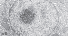
A region within the nucleus, the nucleolus, is where ribosomes begin to be assembled from RNA and proteins.

The contents of the nucleus, aside from the nucleolus, are referred to as the nucleoplasm. Similar to the cytoplasm, the nucleoplasm consists of the liquid content of the nucleus and the insoluble molecules suspended within it.
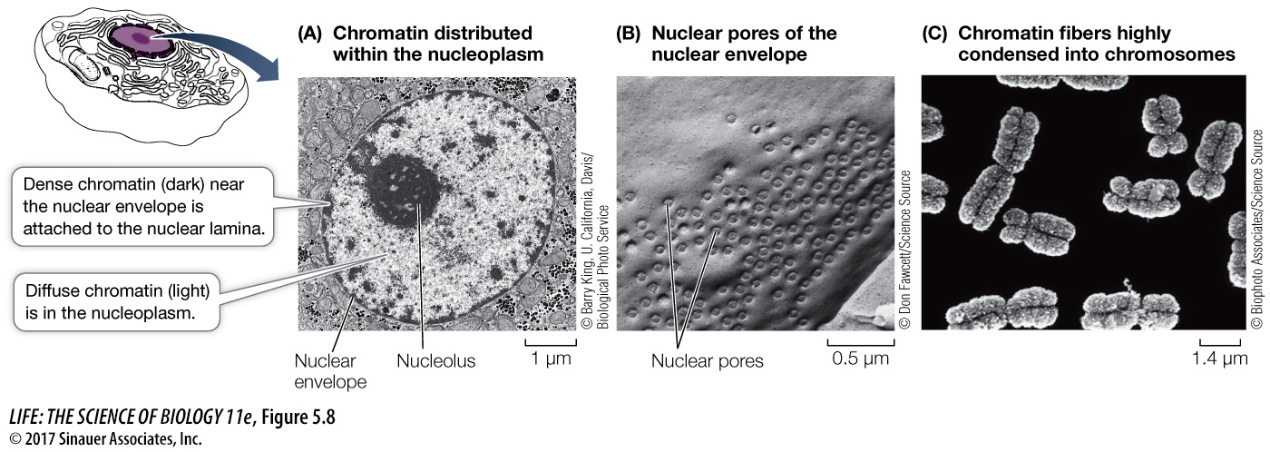
The nucleus is surrounded by an integrated structure composed of two membranes, called the nuclear envelope. This structure separates the genetic material from the cytoplasm. Functionally, it separates DNA transcription (which occurs in the nucleus) from translation (which occurs in the cytoplasm) (see Key Concept 4.1). The two membranes of the nuclear envelope are perforated by thousands of nuclear pores (Figure 5.8B), each measuring approximately 9 nm in diameter, which connect the nucleoplasm with the cytoplasm.
The pores act as “traffic cops,” allowing some molecules to pass into and out of the nucleus and blocking others. This allows the nucleus to regulate its information-
Inside the nucleus, DNA is combined with proteins to form a fibrous complex called chromatin. Chromatin occurs in the form of exceedingly long, thin threads called chromosomes. Different eukaryotic organisms have different numbers of chromosomes (ranging from two in one kind of Australian ant to hundreds in some plants). Prior to cell division, the chromatin becomes tightly compacted and condensed so that the individual chromosomes are visible under a light microscope. This facilitates distribution of the DNA during cell division (Figure 5.8C).
At the inside edge of the nucleus, the chromatin is attached to a protein meshwork, called the nuclear lamina, which is formed by the polymerization of proteins called lamins into long, thin structures called intermediate filaments. The nuclear lamina maintains the shape of the nucleus by its attachment to both the chromatin and the nuclear envelope. On the outside of the nucleus, the outer membrane of the nuclear envelope folds outward into the cytoplasm and is continuous with the membrane of another organelle, the endoplasmic reticulum, which we will discuss next.