The Na+/K+ ATPase Maintains the Intracellular Na+ and K+ Concentrations in Animal Cells
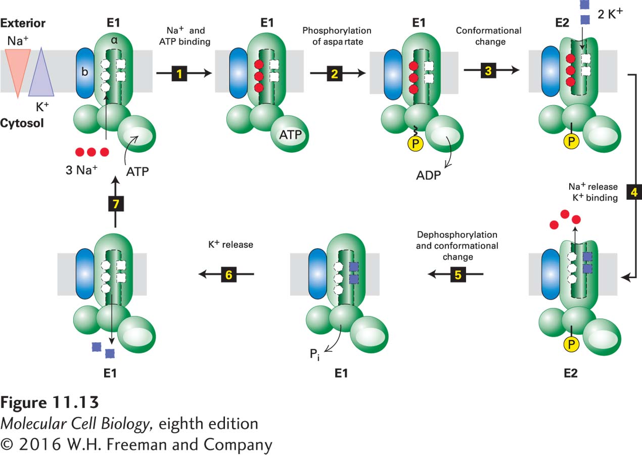
An important P-class pump that is present in the plasma membranes of all animal cells is the Na+/K+ ATPase. This ion pump is a tetramer of subunit composition α2β2 and shares structural homology with the Ca2+ pump (see Figure 11-12). The small, glycosylated β transmembrane polypeptide apparently is not involved directly in ion pumping. During its catalytic cycle, the Na+/K+ ATPase moves three Na+ ions out of and two K+ ions into the cell per ATP molecule hydrolyzed. The mechanism of action of the Na+/K+ ATPase, outlined in Figure 11-13, is similar to that of the muscle SR calcium pump, except that ions are pumped in both directions across the membrane, with each ion moving against its concentration gradient. In its E1 conformation, the Na+/K+ ATPase has three high-affinity Na+-binding sites and two low-affinity K+-binding sites accessible from the cytosolic surface of the protein. The Km for binding of Na+ to these cytosolic sites is 0.6 mM, a value considerably lower than the intracellular Na+ concentration of ~12 mM; as a result, Na+ ions normally fully occupy these sites. Conversely, the affinity of the cytosolic K+-binding sites is low enough that K+ ions, transported inward through the protein, dissociate from E1 and enter the cytosol despite the high intracellular K+ concentration. During the E1 → E2 transition, the three bound Na+ ions gain access to the exoplasmic face, and simultaneously, the affinity of the three Na+-binding sites drops. The three Na+ ions, now bound to low-affinity Na+ sites, dissociate one at a time and enter the extracellular medium despite the high extracellular Na+ concentration. Transition to the E2 conformation also generates two high-affinity K+ sites accessible from the exoplasmic face. Because the Km for K+ binding to these sites (0.2 mM) is lower than the extracellular K+ concentration (4 mM), these sites will fill with K+ ions as the Na+ ions dissociate. Similarly, during the subsequent E2 → E1 transition, the two bound K+ ions are transported inward and then released into the cytosol.
FIGURE 11-13Operational model of the plasma-membrane Na+/K+ ATPase. Only one of the two catalytic α subunits of this P-class pump is depicted. It is not known whether just one or both subunits in a single ATPase molecule transport ions. Ion pumping by the Na+/K+ ATPase involves phosphorylation, dephosphorylation, and conformational changes similar to those in the muscle Ca2+ ATPase (see Figure 11-11). In this case, hydrolysis of the E2–P intermediate powers the E2 → E1 conformational change and concomitant transport of two K+ ions inward. Na+ ions are indicated by red circles; K+ ions, by purple squares; high-energy acyl phosphate bond, by ~P; low-energy phosphoester bond, by –P.
Certain drugs (e.g., ouabain and digoxin) bind to the exoplasmic domain of the plasma-membrane Na+/K+ ATPase and specifically inhibit its ATPase activity. The resulting disruption in the Na+/K+ balance of cells is strong evidence for the critical role of this ion pump in maintaining the normal K+ and Na+ ion concentration gradients. Classic Experiment 11-1 describes the discovery of this important pump, which is required for life.