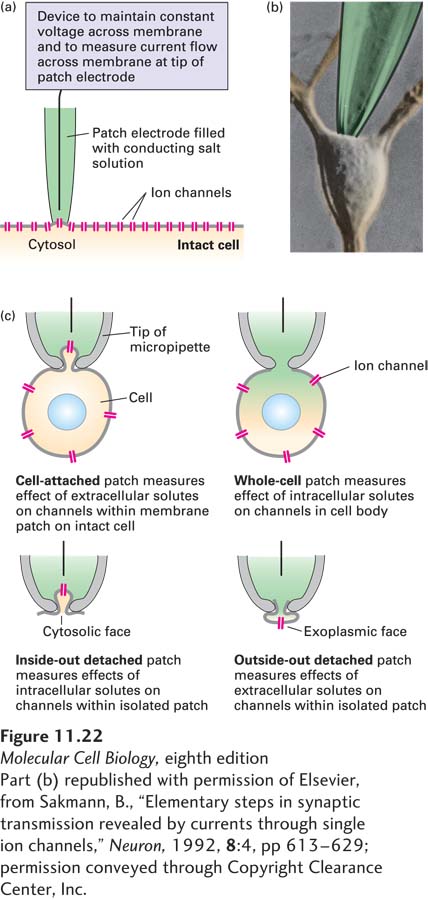
Patch Clamps Permit Measurement of Ion Movements Through Single Channels
Once it was realized that in most cells there are only one or a few ion channels per square micrometer of plasma membrane, it became possible to record ion movements through single ion channels, and to measure the rates at which these channels open and close and conduct specific ions, using a technique known as patch clamping. As illustrated in Figure 11-22, a tiny glass pipette is tightly sealed, using suction, to the surface of a cell; the segment of the plasma membrane within the tip of the pipette will contain only one or a few ion channels. The only current that crosses through the patch of membrane must pass through these channels as ions flow. An electrical recording device detects this ion flow, measured as electric current, through the channels; this ion flow usually occurs in small bursts when a channel is open (Figure 11-23). The electrical device also “clamps” or locks the electric potential across the membrane at a predetermined value (hence the term patch clamping). The inward or outward movement of ions across a patch of membrane is quantified from the amount of electric current needed to maintain the membrane potential at a particular “clamped” value (Figure 11-22a). Four common patch-clamp configurations are used (Figure 11-22c). In cell-attached patch clamping, the pipette forms a tight seal around a small area of membrane to record the current through channels within that small area. In whole-cell patch clamping, the plasma membrane is ruptured so that the glass pipette has access to the cytoplasm to record currents from the entire cell body. In addition, small patches of membrane can be torn away from the cell, with either the inside or the outside of the plasma membrane surface exposed to the extracellular solution. Addition of specific molecules and compounds to the solution within the glass pipette can be used to probe the effects of these reagents on ion channel function.
[Part (b) republished with permission of Elsevier, from Sakmann, B., “Elementary steps in synaptic transmission revealed by currents through single ion channels,” Neuron, 1992, 8:4, pp 613–629; permission conveyed through Copyright Clearance Center, Inc.]
EXPERIMENTAL FIGURE 11-22Current flow through individual ion channels can be measured by patch clamping. (a) Basic experimental arrangement for measuring current flow through individual ion channels in the plasma membrane of a live cell. The patch electrode, filled with a current-conducting saline solution, is applied, with a slight suction, to the plasma membrane. The 0.5-µm-diameter tip covers a region that contains only one or a few ion channels. A recording device measures current flow only through the channel or channels in that region. (b) Photomicrograph of the cell body of a cultured neuron and the tip of a patch pipette touching the plasma membrane. (c) Different patch-clamping configurations. 1 In cell-attached patches, the electrode forms a tight seal around a patch of membrane; it records the effects on channels within this patch of different concentrations of ions and solutes such as extracellular hormones. 2 In whole-cell patch-clamp recording, suction is used to create a hole in the membrane so that the micropipette gains access to the inside of the cell. Reagents can be delivered to the cytoplasm through the micropipette, and their effects on channels in the entire cell body can be recorded. One can also study the effects of different reagents on channels in isolated, detached patches; these are the best configurations for studying the effects on channels of different ion concentrations. 3 In inside-out patches, one can measure the effect of intracellular reagents on channel function by exposing the tip of the micropipette to solutions containing defined reagents. 4Outside-out patches are made from a whole-cell patch configuration by pulling the pipette away from the cell to create a bleb of membrane such that the exoplasmic face of the membrane has access to the solutions to which the tip of the micropipette is exposed. In this way, one can measure the effect of extracellular reagents on channel function. An inside-out patch, in which a cell-attached patch is formed and then the cell pulled away, is used in the experiment in Figure 11-24.
[Part (b) republished with permission of Elsevier, from Sakmann, B., “Elementary steps in synaptic transmission revealed by currents through single ion channels,” Neuron, 1992, 8:4, pp 613–629; permission conveyed through Copyright Clearance Center, Inc.]
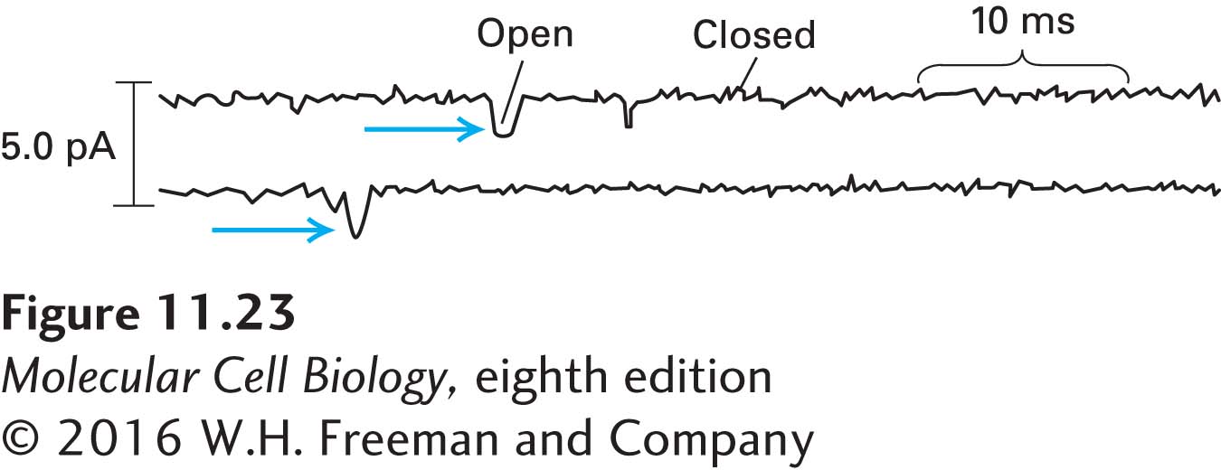
EXPERIMENTAL FIGURE 11-23Ion flux through individual Na+ channels can be calculated from patch-clamp tracings. Two inside-out patches of muscle plasma membrane were clamped at a potential of slightly less than the resting membrane potential. The patch pipette contained NaCl. The transient pulses of electric current (in picoamperes), recorded as large downward deviations (blue arrows), indicate the opening of a Na+ channel and the movement of positive charges (Na+ ions) inward across the membrane. The smaller deviations in current represent background noise. The average current through an open channel is 1.6 pA, or 1.6 × 10⁻¹² amperes. Since 1 ampere = 1 coulomb (C) of charge per second, this current is equivalent to the movement of about 9900 Na+ ions per channel per millisecond: (1.6 × 10⁻¹² C/s)(10−3 s/ms)(6 × 1023 molecules/mol) ÷ 96,500 C/mol. See F. J. Sigworth and E. Neher, 1980, Nature287:447.
Page 501
The tracings in Figure 11-23 illustrate the use of patch clamping to study the properties of voltage-gated Na+ channels (which open in response to changes in membrane potential) in the plasma membrane of muscle cells. As we discuss in Chapter 22, these channels are normally closed in resting muscle cells and open following neuronal stimulation. Patches of muscle-cell plasma membrane, each containing an average of one Na+ channel, were clamped at a predetermined voltage that, in this study, was slightly less than the resting membrane potential. Under these circumstances, transient pulses of positive charges (Na+ ions) cross the membrane from the exoplasmic to the cytosolic face as individual Na+ channels open and then close. Each channel is either fully open or completely closed. From such tracings, it is possible to determine how long a channel is open and the ion flux through it. For the channels measured in Figure 11-23, the flux is about 10 million Na+ ions per channel per second, a typical value for ion channels. Replacement of the NaCl within the patch pipette (corresponding to the outside of the cell) with KCl or choline chloride abolishes current through the channels, confirming that they conduct only Na+ ions, not K+ or other ions.