Chapter Introduction
CHAPTER 22: Cells of the Nervous System

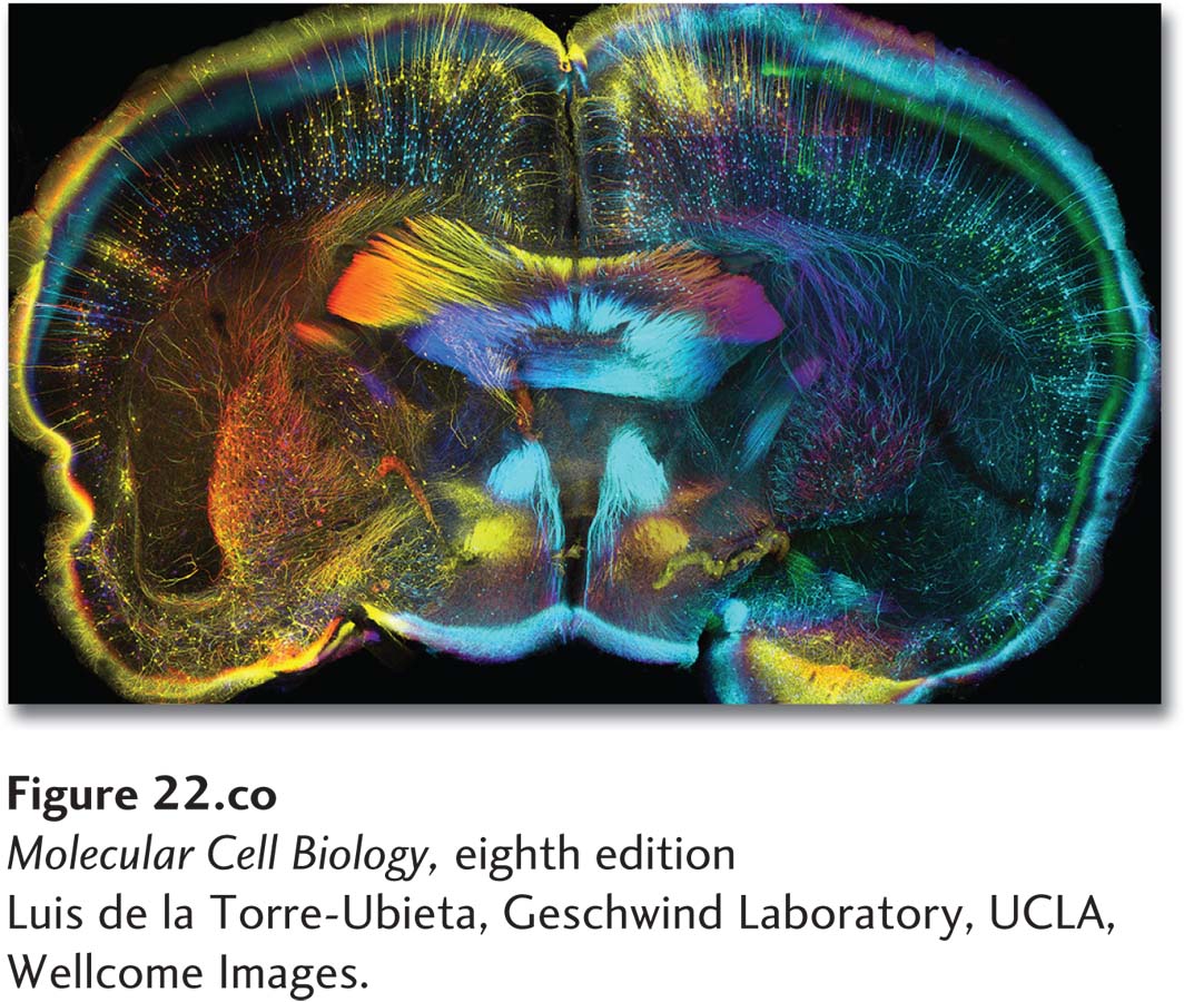
The nervous system regulates all aspects of bodily function and is staggering in its complexity. The 1.3-
Neurons are organized into interconnected units or circuits that have discrete functions. Some circuits sense features of both the external and internal environments of organisms and transmit this information to the brain for processing and storage. Others regulate the contraction of muscles and the secretion of hormones. Yet other circuits regulate cognition, emotion, and innate as well as learned behaviors. In addition to neurons, the nervous system contains glial cells. Historically considered to function simply as support cells for neurons, it is now recognized that glia play active roles in brain function.
The biology of the cells of the nervous system is remarkable on two levels. First, neurons are the most morphologically polarized and compartmentalized cells in the body, and thus pose great challenges to many cell biological processes, from cytoskeletal dynamics and membrane trafficking to signal transduction and gene regulation. Second, individual neurons and glia combine to form exquisitely complex and precise networks or circuits. Neural circuits are not hard wired, but instead the connectivity of neurons changes with experience through a process known as synaptic plasticity, in which experience modifies the strength and number of synaptic connections between neurons. A central focus of modern brain biology is understanding the logic underlying both the formation and the plasticity of neural circuits. While the structure and function of nerve cells is understood in great detail—
Page 1026
The vertebrate nervous system is anatomically divided into the central nervous system, which contains the nerves and glia located inside the brain and spinal cord, and the peripheral nervous system, which contains the nerves and glia located outside the brain and spinal cord. Despite being anatomically separate, the central and peripheral nervous systems are functionally interconnected, with peripheral nerves serving as communication conduits between the brain and the body. The central nervous system itself can be divided into four primary components: the spinal cord, brainstem, cerebellum, and cerebrum. Each region has discrete functions. For example, the spinal cord conducts sensory and motor information from the body to the brain, the brainstem regulates breathing and blood pressure, the cerebellum controls motor function, and the cerebrum processes motor and sensory information, language, learning and memory, and other higher-
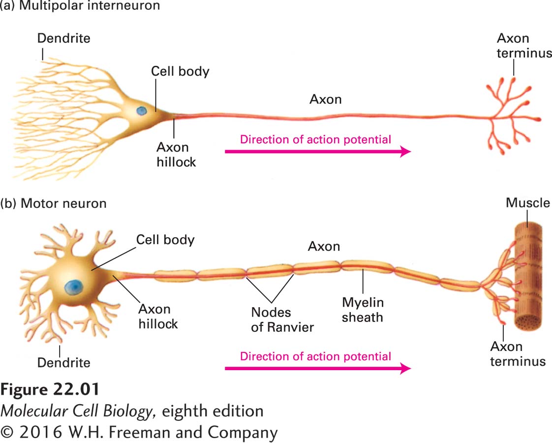
Indeed, despite the multiple types and shapes of neurons that are found in metazoan organisms, all nerve cells share common properties that make them specialized for communicating information using a combination of electrical and chemical signaling. Electrical signals process and conduct information within neurons, which are usually highly polarized cells with extensions whose lengths are orders of magnitude greater than the cell soma (Figure 22-1). The electrical pulses that travel along neurons are called action potentials, and information is encoded as the frequency at which action potentials are fired. Owing to the speed of electrical transmission, neurons are champion signal transducers, much faster than cells that secrete hormones. In contrast to the electrical signals that conduct information within a neuron, chemical signals transmit information between cells, utilizing processes similar to those employed by other types of signaling cells (Chapters 15 and 16).

Taken together, the electrical and chemical signaling of the nervous system allows it to detect external stimuli, integrate and process the information received, relay it to higher brain centers, and generate an appropriate response to the stimulus. For example, sensory neurons have specialized receptors that convert diverse types of stimuli from the environment (e.g., light, touch, sound, odorants) into electrical signals. These electrical signals are then converted into chemical signals that are passed on to other cells called interneurons, which convert the information back into electrical signals. Ultimately the information is transmitted to muscle-
In this chapter we will focus on neurobiology at the cellular and molecular level. We will start by looking at the general architecture of neurons, at how they carry signals, and at how neurons and glia arise from stem cells. Next we will focus on ion flow, channel proteins, and membrane properties: how electrical pulses move rapidly along neurons. Third, we will examine communication between neurons: electrical signals traveling along a cell must be translated into a chemical pulse between cells and then back into an electrical signal in the receiving cells. We will then examine neurons in several sensory tissues, including those that mediate our senses of touch, taste and olfaction. The speed, precision, and integrative power of neural signaling enable the accurate and timely sensory perception of a swiftly changing environment. In the last section, we will turn to the circuits, neurons, and cell biological mechanisms underlying the storage of memories.
A great deal of information about nerve cells has been gleaned from analyses of humans, mice, nematodes, and flies with mutations that affect specific functions of the nervous system. In addition, molecular cloning and structural analysis of key neuronal proteins, such as voltage-