Thylakoid Membranes in Chloroplasts Are the Sites of Photosynthesis in Plants
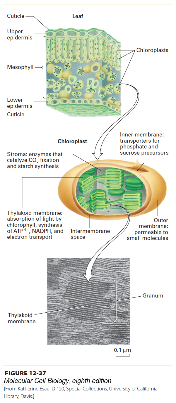
Chloroplasts are lens-shaped organelles with a diameter of approximately 5 µm and a width of approximately 2.5 µm. They contain about 3000 different proteins, 95 percent of which are encoded in the nucleus, made in the cytosol, imported into the organelle, and then transported to their appropriate membrane or space (see Chapter 13). Chloroplasts are bounded by two membranes, which do not contain chlorophyll and do not participate directly in the generation of ATP and NADPH driven by light (Figures 12-37 and 12-38). Like that of mitochondria, the outer membrane of chloroplasts contains porins and thus is permeable to metabolites of small molecular weight. The inner membrane forms a permeability barrier that contains transport proteins for regulating the movement of metabolites into and out of the organelle.
Unlike mitochondria, chloroplasts contain a third membrane—the thylakoid membrane—on which the light-driven generation of ATP and NADPH occurs. The chloroplast thylakoid membrane is believed to constitute a single sheet that forms numerous small, interconnected flattened structures, the thylakoids, which are commonly arranged in stacks termed grana (see Figure 12-37). The spaces within all the thylakoids constitute a single continuous compartment, the thylakoid lumen (see Figure 12-38). The thylakoid membrane contains a number of integral membrane proteins to which are bound several important prosthetic groups and light-absorbing pigments, most notably chlorophylls. Starch synthesis and storage occurs in the stroma, the aqueous compartment between the thylakoid membrane and the inner membrane. In photosynthetic bacteria, extensive invaginations of the plasma membrane form a set of internal membranes, also termed thylakoid membranes, where photosynthesis occurs.
[From Katherine Esau, D-120, Special Collections, University of California Library, Davis.]
FIGURE 12-37Structure of leaf and chloroplast. Like mitochondria, plant chloroplasts are bounded by two membranes separated by an intermembrane space. Photosynthesis occurs on a third membrane, the thylakoid membrane, which is surrounded by the inner membrane and forms a series of flattened vesicles (thylakoids) that enclose a single interconnected luminal space. The green color of plants is due to the green color of chlorophyll, all of which is located within the thylakoid membrane. A granum is a stack of adjacent thylakoids. The stroma is the space between the inner membrane and the thylakoids.
[From Katherine Esau, D-120, Special Collections, University of California Library, Davis.]
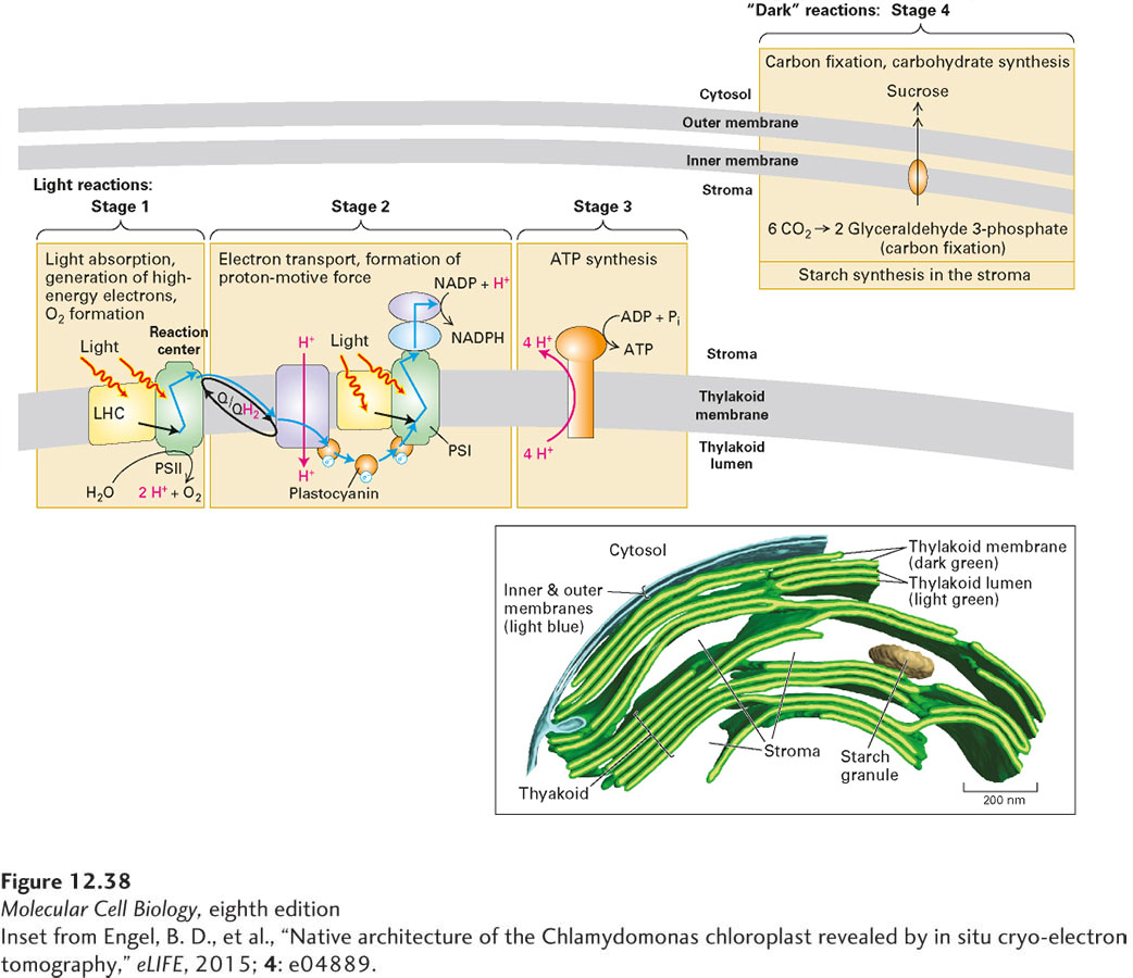
[Inset from Engel, B. D., et al., “Native architecture of the Chlamydomonas chloroplast revealed by in situ cryo-electron tomography,” eLIFE, 2015; 4: e04889.]
FIGURE 12-38Overview of the four stages of photosynthesis. In stage 1, light is absorbed by light-harvesting complexes (LHCs) and the reaction center of photosystem II (PSII). The LHCs transfer the absorbed energy to the reaction centers, which use it, or the energy absorbed directly from a photon, to oxidize water to molecular oxygen and generate high-energy electrons (electron paths shown by blue arrows). In stage 2, these electrons move down an electron-transport chain, which uses either lipid-soluble (Q/QH2) or water-soluble (plastocyanin, PC) electron carriers to shuttle electrons between multiple protein complexes. As electrons move down the chain, they release energy that the complexes use to generate a proton-motive force and, after additional energy is introduced by absorption of light in photosystem I (PSI), to synthesize the high-energy electron carrier NADPH. In stage 3, flow of protons down their concentration and voltage gradient through the F0F1 ATP synthase drives ATP synthesis. Stages 1–3 in plants take place in the thylakoid membrane of the chloroplast. In stage 4, in the chloroplast stroma, the energy stored in NADPH and ATP is used to incorporate CO2 into the three-carbon molecule glyceraldehyde 3-phosphate, the first step in a process known as carbon fixation. These molecules are then transported to the cytosol of the cell for conversion to hexose sugars in the form of sucrose. Glyceraldehyde 3-phosphate is also used to make starch within the chloroplast. Inset: Three-dimensional reconstruction from cryoelectron tomography of a chloroplast in the unicellular green alga Chlamydomonas reinhardtii, showing thylakoid membranes (dark green), thylakoid lumen (light green), inner and outer membranes (blue), and one small starch granule (tan).
[Inset from Engel, B. D., et al., “Native architecture of the Chlamydomonas chloroplast revealed by in situ cryo-electron tomography,” eLIFE, 2015; 4: e04889.]