Recruitment of PI-3 Kinase to Activated Receptors Leads to Synthesis of Three Phosphorylated Phosphatidylinositols
Besides the IP3/DAG pathway, many activated RTKs and cytokine receptors initiate another phosphoinositide pathway by recruiting the enzyme phosphatidylinositol-3 kinase (PI-3 kinase) to the membrane. PI-3 kinase is recruited to the cytosolic surface of the plasma membrane by binding of its SH2 domain to phosphotyrosines on the cytosolic domain of many activated RTKs and cytokine receptors. This recruitment positions the catalytic domain of PI-3 kinase near its phosphoinositide substrates on the cytosolic face of the plasma membrane. Unlike kinases we have encountered earlier that phosphorylate proteins, PI-3 kinase adds a phosphate to the 3′ carbon in one of two separate phosphatidylinositol substrates, leading to formation of two phosphatidylinositol 3-phosphates: phosphatidylinositol 3,4-bisphosphate [PI(3,4)P2] or phosphatidylinositol 3,4,5-trisphosphate [PI(3,4,5)P3] (Figure 16-28). By acting as docking sites for various signal-transducing proteins, these membrane-bound PI 3-phosphate products of the PI-3 kinase reactions in turn transduce signals downstream in several important pathways.
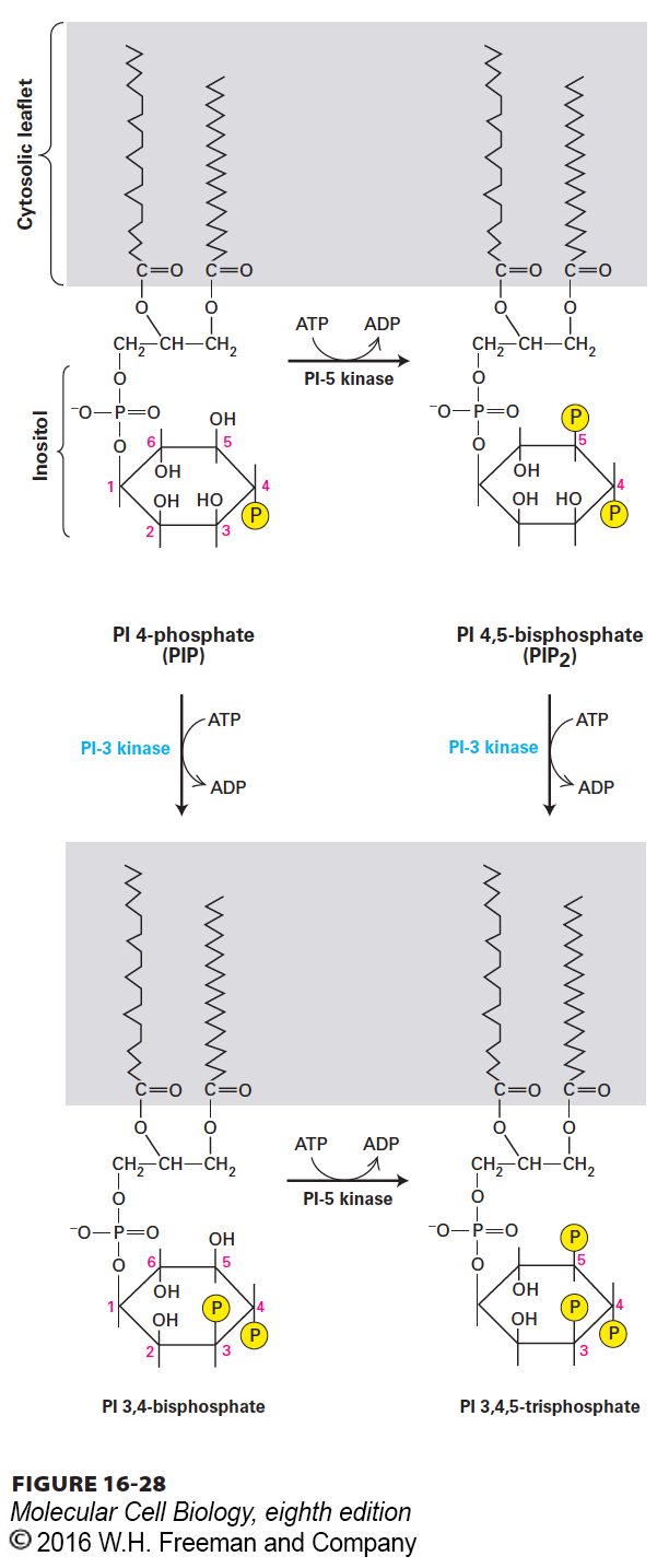
FIGURE 16-28Generation of phosphatidylinositol 3-phosphates. The enzyme phosphatidylinositol-3 kinase (PI-3 kinase) is recruited to the membrane by many activated receptor tyrosine kinases (RTKs) and cytokine receptors. The 3-phosphate added by this enzyme, to yield PI(3,4)P2 or PI(3,4,5)P3, is a binding site for various signal-transducing proteins, such as the PH domain of protein kinase B. Phosphatidylinositol 4,5-bisphosphate is also the substrate of phospholipase C (see Figure 15-33). See L. Rameh and L. C. Cantley, 1999, J. Biol. Chem.274:8347.
In some cells, this PI-3 kinase pathway can trigger cell division and prevent apoptosis, thus ensuring cell survival. In other cells, this pathway induces specific changes in cell metabolism. PI-3 kinase was first identified in studies of the polyoma virus, a DNA virus that transforms certain mammalian cells. Transformation requires several virally encoded oncoproteins, including one termed “middle T.” In an attempt to discover how middle T functions, investigators found PI-3 kinase in partially purified preparations of middle T, which suggested a specific interaction between the two. Then they set out to determine how PI-3 kinase might affect cell behavior.
Page 750
When an inactive, dominant-negative version of PI-3 kinase was expressed in polyoma virus–transformed cells, it inhibited the uncontrolled cell proliferation characteristic of virus-transformed cells. This finding suggested that the normal kinase is important in certain signaling pathways essential for cell proliferation or for the prevention of programmed cell death (apoptosis; see Chapter 21). Subsequent work showed that PI-3 kinases participate in many signaling pathways related to cell growth and apoptosis. Of the nine PI-3 kinase homologs encoded by the human genome, the best characterized contains a p110 subunit with catalytic activity and a p85 subunit with an SH2 phosphotyrosine-binding domain.