Actin and Myosin II Form Contractile Bundles in Nonmuscle Cells
In skeletal muscle, as we have seen, actin thin filaments and myosin II thick filaments assemble into contractile structures. Nonmuscle cells contain several types of related contractile bundles composed of actin and myosin II filaments, which are similar to skeletal muscle fibers but much less organized. Moreover, they lack the troponin regulatory system and are instead regulated by myosin phosphorylation, as we discuss below.
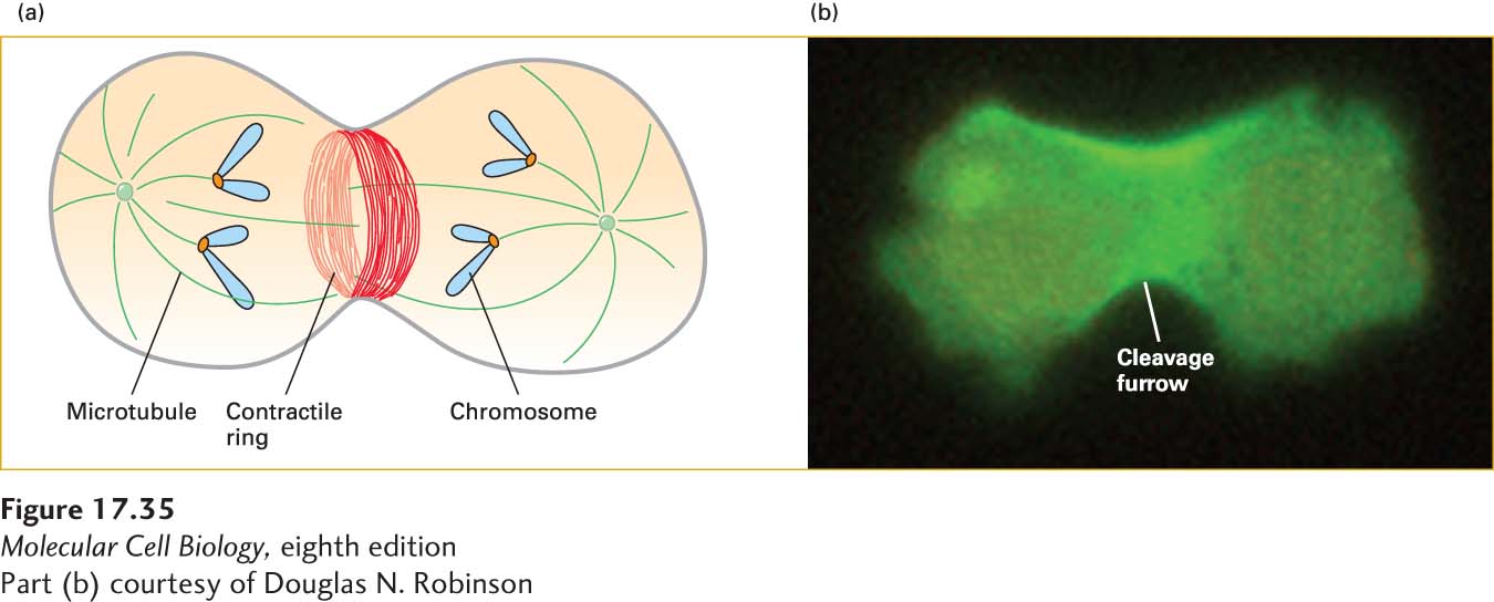
In epithelial cells, contractile bundles are most commonly found as an adherens belt, also known as the circumferential belt, that encircles the inner surface of the cell at the level of the adherens junction (see Figure 17-4a). These bundles are important in maintaining the integrity of the epithelium (discussed in Chapter 20). Stress fibers, which are seen along the lower surfaces of cells cultured on artificial (glass or plastic) surfaces or on extracellular matrices, are a second type of contractile bundle (see Figure 17-4a, c) important in cell adhesion, especially on deformable substrata. The ends of stress fibers terminate at integrin-containing focal adhesions, special structures that attach a cell to the underlying substratum (see Figure 17-40 below and Chapter 20). Circumferential belts and stress fibers contain several proteins found in the contractile apparatus of smooth muscle and exhibit some organizational features resembling those of muscle sarcomeres. A third type of contractile bundle, referred to as a contractile ring, is a transient structure in animal cells that assembles at the equator of a dividing cell, encircling the cell midway between the poles of the mitotic spindle (Figure 17-35a). As the ring contracts, pulling the plasma membrane in, the cytoplasm is divided and eventually pinched into two parts in a process known as cytokinesis, giving rise to two daughter cells. Dividing cells stained with antibodies against myosin I and myosin II show that myosin II is localized to the contractile ring, whereas myosin I is at the distal regions, where it links the cell cortex to the plasma membrane (Figure 17-35b). Cells from which the gene encoding the heavy chain of myosin II has been deleted are unable to undergo cytokinesis. Instead, these cells form a multinucleated syncytium because cytokinesis, but not nuclear division, is inhibited.
[Part (b) courtesy of Douglas N. Robinson]
EXPERIMENTAL FIGURE 17-35Fluorescent antibodies reveal the localization of myosin I and myosin II during cytokinesis. (a) Diagram of a cell going through cytokinesis, showing the mitotic spindle (microtubules green, chromosomes blue) and the contractile ring with actin filaments (red). (b) Fluorescence micrograph of a Dictyostelium amoeba expressing GFP-myosin-II reveals myosin-II enrichment in the cleavage furrow cortex, as the cell pinches into two during cytokinesis.