CHAPTER18:Cell Organization and Movement II: Microtubules and Intermediate Filaments
[Reprinted by permission from Macmillan Publishers Ltd: A. Khodjakov, “Olympus/Nature competition: A 1, 2, 3 in light microscopy,” Nature 408, 423-424. Courtesy Alexey Khodjakov.]
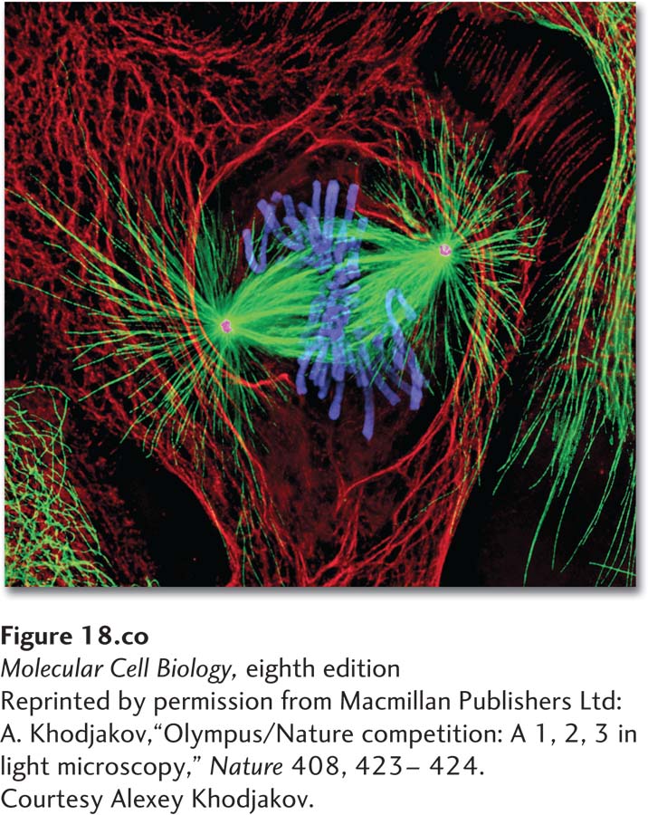
Newt lung cell in mitosis stained for centrosomes (magenta), microtubules (green), chromosomes (blue), and keratin intermediate filaments (red).
[Reprinted by permission from Macmillan Publishers Ltd: A. Khodjakov, “Olympus/Nature competition: A 1, 2, 3 in light microscopy,” Nature 408, 423-424. Courtesy Alexey Khodjakov.]
As we learned in the previous chapter, three types of filaments make up the animal-cell cytoskeleton: microfilaments, microtubules, and intermediate filaments. Why have these three distinct types of filaments evolved? It seems likely that their physical properties are suited to different functions. In Chapter 17, we described how actin microfilaments are often cross-linked into networks or bundles to form flexible and dynamic structures and to serve as tracks for the many different classes of myosin motors. Similarly, microtubules are stiff tubes that can exist as a single structure, extending up to 20 µm in cells, or in bundled arrangements such as those seen in specialized cell-surface structures like cilia and flagella. As a consequence of their tubular design, microtubules are able to generate pulling and pushing forces without buckling, a property that allows single tubules to extend long distances within a cell and bundles to slide past each other, as occurs in flagella and in the mitotic spindle. Microtubules’ ability to extend long distances in the cell, together with their intrinsic polarity, is exploited by microtubule-dependent motors, which use microtubules as tracks for long-range transport of organelles. Microtubules can be highly dynamic—assembling and disassembling rapidly from their ends—and can thus provide the cell with the flexibility to alter microtubule organization as needed.
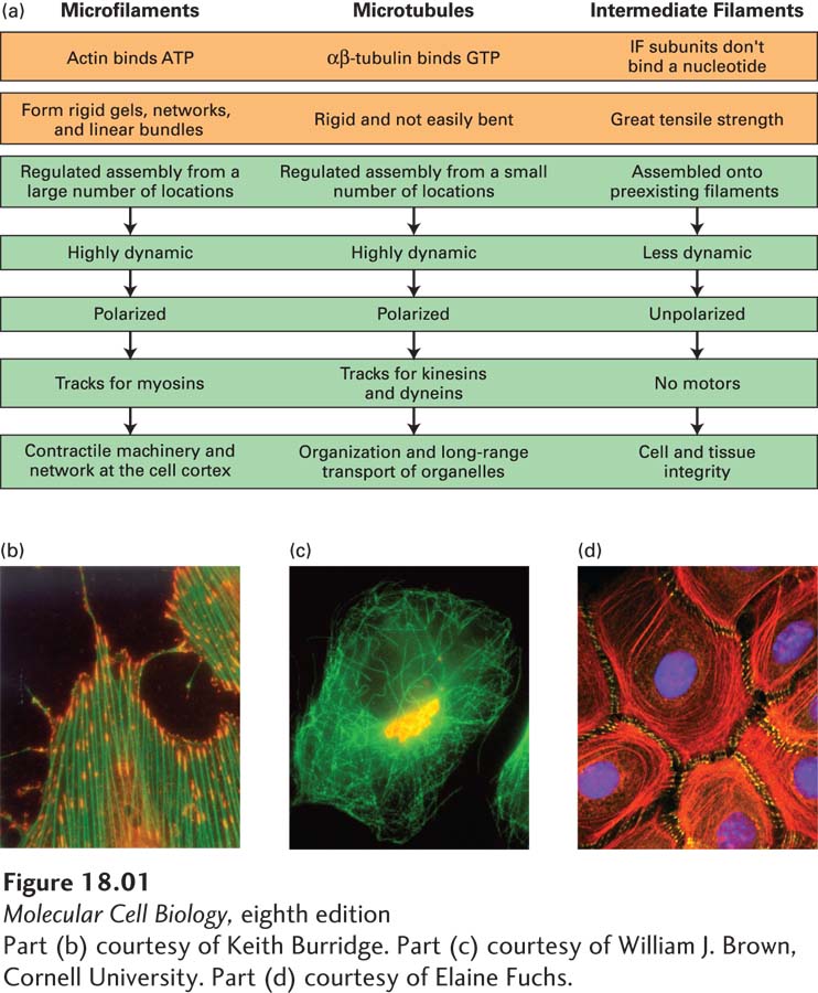
In contrast to microfilaments and microtubules, intermediate filaments have great tensile strength and have evolved to withstand much larger stresses and strains. With properties akin to strong molecular ropes, they are ideally suited to endow both cells and tissues with structural integrity and to contribute to cellular organization. Intermediate filaments do not have an intrinsic polarity as microfilaments and microtubules do, so it is not surprising that there are no known motor proteins that use intermediate filaments as tracks. Although we discuss microtubules and intermediate filaments together in this chapter—and although their localization in the cytosol can look superficially quite similar—we will see that their dynamics and functions are very different. A summary of the similarities and differences among these three filament systems is shown in Figure 18-1.
Page 822
[Part (b) courtesy of Keith Burridge. Part (c) courtesy of William J. Brown, Cornell University. Part (d) courtesy of Elaine Fuchs.]
FIGURE 18-1Overview of the physical properties and functions of the three filament systems in animal cells. (a) Biophysical and biochemical properties (orange) and biological properties (green) are shown for each filament type. The micrographs (b–d) show examples of each filament type in a particular cellular context, but note that microtubules also make up other structures, and that intermediate filaments also line the inner surface of the nucleus. (b) Cultured cells stained for actin (green) and sites of actin attachment to the substratum (orange). (c) Localization of microtubules (green) and the Golgi complex (yellow). Notice the central location of the Golgi complex, which is collected there by transport along microtubules. (d) Localization of cytokeratins (red), a type of intermediate filament, and a component of desmosomes (yellow) in epithelial cells. Cytokeratins from individual cells are attached to each other through the desmosomes.
[Part (b) courtesy of Keith Burridge. Part (c) courtesy of William J. Brown, Cornell University. Part (d) courtesy of Elaine Fuchs.]
This chapter covers five main topics. First, we discuss the structure and dynamics of microtubules and their motor proteins. Second, we examine how microtubules and their motors contribute to the movement of cilia and flagella. Third, we discuss the role of microtubules in the mitotic spindle—a molecular machine that has evolved to accurately segregate duplicated chromosomes. Fourth, we explore the roles of the different classes of intermediate filaments that provide structure to the nuclear envelope as well as strength and organization to cells and tissues. Although we consider microtubules, microfilaments, and intermediate filaments individually, the three filament systems do not act independently of one another, and we consider some examples of this interdependence in the last section of the chapter.