Microtubules Are Stabilized by Side-Binding Proteins
Several different classes of proteins stabilize microtubules, many of which show cell-type-specific expression. Among the best-studied MAPs are members of the tau family of proteins, which includes tau itself as well as proteins called MAP2 and MAP4. Tau and MAP2 are neuronal proteins, while MAP4 is expressed by other cell types and is generally not present in neurons. These proteins have a modular design with two key domains. The first domain consists of a positively charged 18-residue sequence, repeated three to four times, that binds to the negatively charged microtubule surface. The second domain projects outward at a right angle from the microtubule (Figure 18-13). Tau proteins are believed to stabilize microtubules and also to act as spacers between them. MAP2 is found only in dendrites of neurons, where it forms fibrous cross-bridges between microtubules and links microtubules to intermediate filaments. Tau, which is much smaller than most other MAPs, is present in both axons and dendrites. The basis for this specificity is still a mystery.
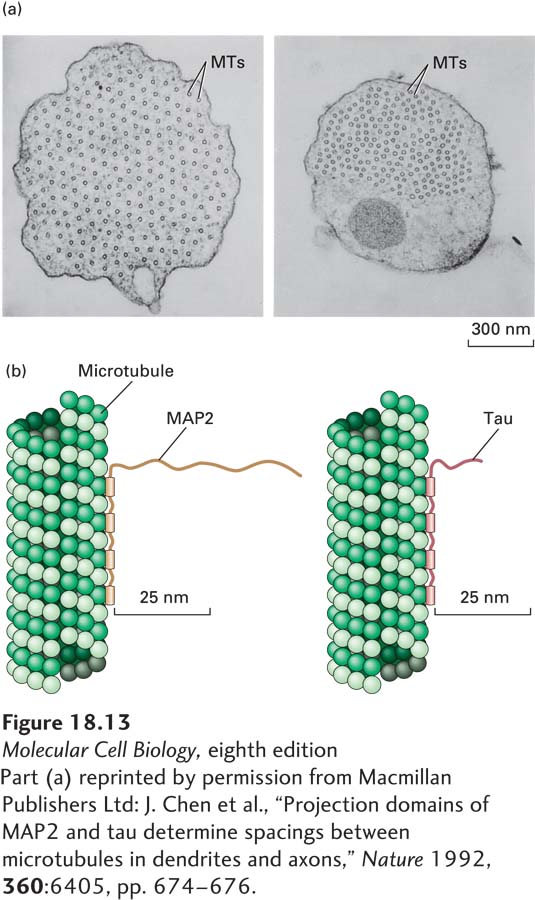
[Part (a) reprinted by permission from Macmillan Publishers Ltd: J. Chen et al., “Projection domains of MAP2 and tau determine spacings between microtubules in dendrites and axons,” Nature 1992, 360:6405, pp. 674-676.]
EXPERIMENTAL FIGURE 18-13Spacing of microtubules depends on the length of the projection domains of microtubule-associated proteins. (a) Insect cells transfected to express MAP2, which has a long arm, or to express tau protein, which has a short arm, grow long axon-like processes. These electron micrographs show cross sections through the processes induced by the expression of MAP2 (left) and tau (right) in transfected cells. Note that the spacing between microtubules (MTs) in MAP2-containing cells is greater than in tau-containing cells. Both cell types contain approximately the same number of microtubules, but the effect of MAP2 is to enlarge the caliber of the axon-like process. (b) Diagrams of association between microtubules and MAPs. Note the difference between the lengths of the projection domains in MAP2 and in tau.
[Part (a) reprinted by permission from Macmillan Publishers Ltd: J. Chen et al., “Projection domains of MAP2 and tau determine spacings between microtubules in dendrites and axons,” Nature 1992, 360:6405, pp. 674-676.]
Page 831
When stabilizing MAPs coat the outer wall of a microtubule, they can increase the growth rate of microtubules or reduce catastrophe frequency. In many cases, the activity of the MAPs is regulated by the reversible phosphorylation of their projection domains. Phosphorylated MAPs are unable to bind to microtubules; thus phosphorylation promotes microtubule disassembly. For example, microtubule-affinity-regulating kinase (MARK/Par-1) is a key modulator of tau proteins. Some MAPs, including MAP4, are also phosphorylated by a cyclin-dependent kinase (CDK) that plays a major role in controlling the activities of proteins in the course of the cell cycle (see Chapter 19).