Somatic Cells Can Generate iPS Cells

Because of the inefficiency of somatic-cell nuclear transfer, it remained unclear whether all types of somatic mammalian cells retained an intact genome and whether they could be induced to dedifferentiate into an ES cell–like state. Shinya Yamanaka used retrovirus vectors to express a wide variety of transcription factors, singly and in combination, in cultured fibroblast cells. Remarkably, he found that both human and mouse fibroblasts could be reprogrammed to a pluripotent state, called an induced pluripotent stem-cell state, similar to that of an embryonic stem cell, by transformation with retroviruses encoding just four proteins: KLF4, Sox2, Oct4, and Myc. Note that two of these, Sox2 and Oct4, are two of the master transcription factors expressed in ES cells, as discussed previously. In addition to fibroblasts, keratinocytes (skin-forming cells) and other types of differentiated cells have been reprogrammed to iPS cells. Like ES cells, single mouse iPS cells can be experimentally introduced into a blastocyst and form all of the tissues of a mouse, including germ cells, attesting to the fact that somatic cells can indeed be reprogrammed to an embryonic pluripotent state.

Several other transcription factors, and even certain small organic molecules, can replace the Oct4 gene in the Yamanaka reprogramming “cocktail.” Subsequent analysis led to the discovery that each of these factors directly activates transcription of the endogenous (cellular) Oct4 gene, leading to induction of pluripotency. Thus it was hypothesized that, over time, forced expression of transcription-factor genes activates expression of many cellular genes, including those encoding Oct4 and other pluripotency proteins; over the course of several weeks, this activation reprograms the somatic cells to an ES-like state. To experimentally establish the point that activation of endogenous genes leads to reprogramming to an ES-like state, cultured keratinocytes were repeatedly transfected with synthetic mRNAs encoding the four canonical Yamanaka transcription factors, KLF4, Sox2, Oct4, and Myc. These cultured cells generated normal iPS cells that had no trace of any of the exogenously added mRNAs, attesting to the reprogramming of keratinocytes into iPS cells by inducing expression of only normal cellular genes.

In fibroblasts, the chromatin of most pluripotency-associated genes is inaccessible to transcription-factor binding, primarily due to the repressive histone H3 lysine 9 trimethylation (H3K9Me3) mark. Among the genes activated by Oct4 are two that encode H3K9 demethylases, which remove these repressive chromatin marks and, over time, result in activation of pluripotency genes. Consistent with this notion, expression of these H3K9 demethylases increases during reprogramming, and their knockdown inhibits efficient iPS-cell generation. Indeed, reprogramming involves major changes in epigenetic modifications, including DNA methylation and several other types of histone modifications that serve to repress or allow potential activation of hundreds of genes.

984

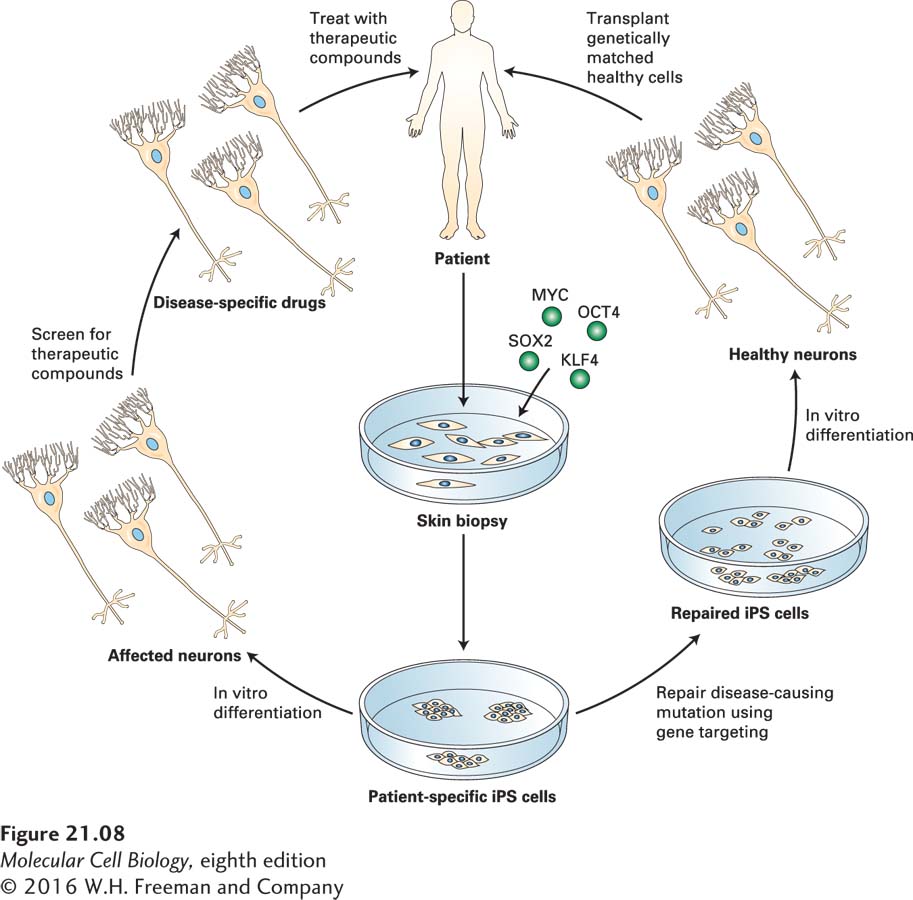
Because iPS cells can be derived from somatic cells of patients with difficult-to-understand diseases, they have already proved invaluable in uncovering the molecular and cellular basis of several afflictions (Figure 21-8). Consider amyotrophic lateral sclerosis (ALS), often called Lou Gehrig’s disease, a fatal disease in which the motor neurons that connect the spinal cord to the muscles of the body progressively die off, causing muscle weakness and death, limb paralysis, and ultimately death due to respiratory failure. There is no cure.

image
FIGURE 21-8 Medical applications of iPS cells. In this example, the patient has a neurodegenerative disorder caused by abnormalities in certain nerve cells (neurons). Patient-specific iPS cells—in this case derived by recombinant expression of transcription factors in cells isolated from a skin biopsy—can be used in one of two ways. In cases in which the disease-causing mutation is known (for example, familial Parkinson’s disease), gene targeting could be used to repair the DNA sequence (right). The gene-corrected patient-specific iPS cells would then undergo directed differentiation into the affected neuronal subtype (for example, midbrain dopaminergic neurons) and be transplanted into the patient’s brain (to engraft the nigrostriatal axis). Alternatively, directed differentiation of the patient-specific iPS cells into the affected neuronal subtype (left) will allow the patient’s disease to be modeled in vitro, and potential drugs can be screened, aiding in the discovery of novel therapeutic compounds. See D. A. Robinton and G. Q. Daley, 2012, Nature 481:295.

In approximately 10 percent of patients, the disease is dominantly inherited (familial ALS), but in 90 percent of patients, there is no apparent genetic linkage (sporadic ALS). An analysis of the underlying causes of the disease at a molecular and cellular level was impossible for many years because one cannot simply extract neurons or the surrounding glial cells from living humans and analyze or culture them.

In about 20 percent of patients with familial ALS, there is a point mutation in the gene SOD1, encoding Cu/Zn superoxide dismutase 1; the mutant SOD1 protein forms aggregates that can damage cells. About 40 percent of patients with familial ALS and 10 percent of patients with the noninherited form have a mutation in the C9ORF72 gene (of unknown function; called chromosome 9 open reading frame 72). This mutation also often occurs in people with frontotemporal dementia, the second most common form of dementia after Alzheimer’s disease, explaining why some people develop both diseases simultaneously. The mRNA transcribed from normal human C9ORF72 genes has up to 30 repeats of the hexanucleotide GGGGCC, but mutant ALS-causing genes can have up to thousands of these repeats.

985

In several studies, iPS cells derived from the skin cells of elderly patients with these and other familial and sporadic forms of the disease were successfully differentiated in culture to form motor neurons; this success demonstrated the feasibility of leveraging the self-renewal of iPS cells to generate a potentially limitless supply of the cells specifically affected by ALS. One study showed that motor neurons bearing several types of ALS mutations were hyperexcitable, generating more of the electrical signals called action potentials (see Chapter 22) than normal. This excess excitability also caused the neurons to make more errors in protein folding and accumulate misfolded proteins, leading to aberrant cell function. In iPS-derived neurons from patients with the C9ORF72 mutation, the RNAs containing the large numbers of repeating GGGGCC sequences were in aggregates, bound to multiple RNA-binding proteins important for normal cell functions; this binding prevented these proteins from catalyzing key steps in the production of other cellular mRNAs. Overall, the C9ORF72 mutation made the motor neurons produce abnormal amounts of many other cellular RNAs and made the cells very sensitive to stress.

In a separate study to dissect the molecular cause of ALS, motor neurons were generated from human ES or iPS cells and cultured with primary human astrocytes, a type of glial cell that surrounds neurons and regulates several of their functions (see Figure 22-17). Many of the motor neurons died if the astrocytes expressed the mutant form of SOD1, but not if they expressed the wild-type form, suggesting that at least in this familial form of ALS, the defective cells are both astrocytes and motor neurons. Indeed, astrocytes expressing the mutant form of SOD1 secreted protein factors that were toxic to adjacent motor neurons.

986

In these and several other studies, researchers screened thousands of small organic molecules, including many approved as drugs for treatment of other unrelated diseases, for those that could reverse the abnormalities in the ALS iPS cell–derived motor neurons. Several were identified and are in clinical trials to see if they can slow or stop the devastating effects of ALS. In any case, these experiments illustrate the value of iPS and ES cells in generating cell culture models of many types of difficult-to-study human diseases that can be used to screen for drugs that could treat many as yet untreatable afflictions.