Cells and Tissues Are Cut into Thin Sections for Viewing by Electron Microscopy
[Keith R. Porter Archive, University of Maryland, Baltimore County.]
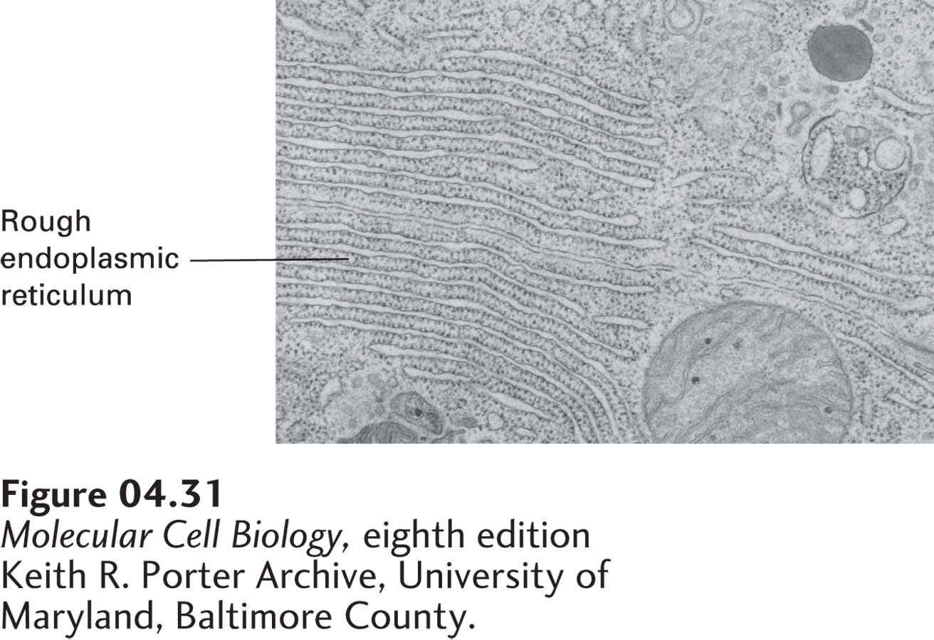
FIGURE 4-31Example of a thin section viewed by transmission electron microscopy. Section through a pancreatic cell showing the extensive rough endoplasmic reticulum involved in the synthesis and secretion of digestive enzymes.
[Keith R. Porter Archive, University of Maryland, Baltimore County.]
Single cells and pieces of tissue are too thick to be viewed directly in the standard transmission electron microscope. To overcome this problem, methods were developed to prepare and cut thin sections of cells and tissues. When these sections were examined in the electron microscope, the organization, beauty, and complexity of the cell interior was revealed and led to a revolution in cell biology—for the first time, new organelles and the first glimpses of the cytoskeleton were seen.
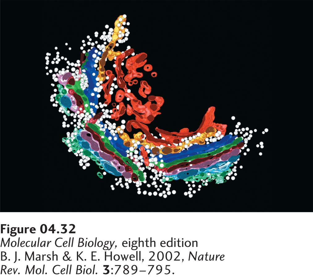
To prepare thin sections, it is necessary to chemically fix the sample, dehydrate it, impregnate it with a liquid plastic that hardens (similar to Plexiglas), and then cut sections of about 5 to 100 nm in thickness. For structures to be seen, the sample has to be stained with heavy metals such as uranium and lead salts, which can be done either before embedding in the plastic or after sections are cut. Examples of cells and tissues viewed by thin-section electron microscopy appear here and throughout this book (Figure 4-31). It is important to realize that the images obtained represent just a thin slice through a cell, so to get a three-dimensional view, it is necessary to cut serial sections through the sample and reconstruct the sample from a series of sequential images (Figure 4-32).
Page 159
[B. J. Marsh & K. E. Howell, 2002, Nature Rev. Mol. Cell Biol.3:789–795.]
FIGURE 4-32Model of the Golgi complex based on three-dimensional reconstruction of electron microscopy images. Transport vesicles (white spheres) that have budded off the rough ER fuse with the cis membranes (light blue) of the Golgi complex. By mechanisms described in Chapter 14, proteins move from the cis region to the medial region and finally to the trans region of the Golgi complex. Eventually, vesicles bud off the trans-Golgi membranes (orange and red); some move to the cell surface and others move to lysosomes. The Golgi complex, like the rough endoplasmic reticulum, is especially prominent in secretory cells.
[B. J. Marsh & K. E. Howell, 2002, Nature Rev. Mol. Cell Biol.3:789–795.]