Chapter Introduction

631

CHAPTER 14

Vesicular Traffic, Secretion, and Endocytosis

image
Schematic representation of the formation of a clathrin-coated vesicle. The process starts with the recruitment of AP2 complexes (green) to the inner surface of the plasma membrane, which in turn capture a three-legged clathrin triskelion. The curvature of the underlying membrane increases as assembly of the lattice proceeds by the recruitment of additional AP2 and clathrin triskelions, until scission results in a coated vesicle formation.
[Courtesy of Ema Cocucci, Janet Iwasa, and Tom Kirchhausen.]

OUTLINE

14.1 Techniques for Studying the Secretory Pathway

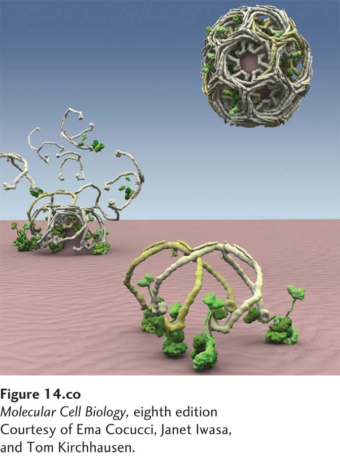
14.2 Molecular Mechanisms of Vesicle Budding and Fusion

14.3 Early Stages of the Secretory Pathway

14.4 Later Stages of the Secretory Pathway

14.5 Receptor-Mediated Endocytosis

14.6 Directing Membrane Proteins and Cytosolic Materials to the Lysosome

In the previous chapter, we explored how proteins are targeted to and translocated across the membranes of several different intracellular organelles, including the endoplasmic reticulum, mitochondria, chloroplasts, peroxisomes, and the nucleus. In this chapter, we turn our attention to the secretory pathway and the mechanisms of vesicular traffic that allow proteins to be secreted from the cell or delivered to the plasma membrane and the lysosomes. We also discuss the related processes of endocytosis and autophagy, which deliver proteins and small molecules either from outside the cell or from the cytoplasm to the interior of the lysosome for degradation.

The secretory pathway is so named because it was initially studied in dedicated secretory cells that produce and secrete large quantities of proteins such as insulin or digestive enzymes to the outside of the cell. It was later discovered that the same pathway used for extracellular secretion of proteins is used to distribute all soluble and membrane proteins that enter the ER to their final destinations at the cell surface or in the lysosomes. Proteins delivered to the plasma membrane include cell-surface receptors, transporters for nutrient uptake, and ion channels that maintain the proper ionic and electrochemical balance across the plasma membrane. Such membrane proteins, once they reach the plasma membrane, remain embedded within it. Soluble secreted proteins also follow the secretory pathway to the cell surface, but instead of remaining embedded in the membrane, they are released into the aqueous extracellular environment. Examples of secreted proteins are digestive enzymes, peptide hormones, serum proteins, and collagen. The lysosome, as described in Chapter 4, is the organelle with an acidic interior that is used for degradation of unneeded proteins and storage of small molecules such as amino acids. Accordingly, the types of proteins delivered to the lysosomal membrane include subunits of the V-class proton pump that pumps H+ from the cytosol into the acidic lumen of the lysosome, as well as transporters that release small molecules stored in the lysosome into the cytoplasm. Soluble proteins delivered by this pathway include lysosomal digestive enzymes such as proteases, glycosidases, phosphatases, and lipases.

632

In contrast to the secretory pathway, which allows proteins to be targeted to the cell surface, the endocytic pathway is used to take up substances from the cell surface and move them into the interior of the cell. The endocytic pathway can selectively remove proteins from the plasma membrane and thus has a part in regulating the protein composition of the plasma membrane. In addition, the endocytic pathway is used to ingest certain nutrients that are too large to be transported across the plasma membrane by one of the transport mechanisms discussed in Chapter 11. For example, the endocytic pathway is used in the uptake of cholesterol carried in LDL particles and of iron atoms carried by the iron-binding protein transferrin. In addition, the endocytic pathway can be used to remove receptor proteins from the cell surface as a way to down-regulate their activity.

A single unifying principle governs all protein trafficking in the secretory and endocytic pathways: transport of membrane and soluble proteins from one membrane-bounded compartment to another is mediated by transport vesicles that collect cargo proteins in buds arising from the membrane of one compartment and then deliver these cargo proteins to the next compartment by fusing with the membrane of that compartment. Importantly, as transport vesicles bud from one membrane and fuse with the next, the same face of the membrane remains oriented toward the cytosol. Therefore, once a protein has been inserted into the membrane or the lumen of the ER, that protein can be carried along the secretory pathway, moving from one organelle to the next without being translocated across another membrane or altering its orientation within the membrane. Similarly, the endocytic pathway uses vesicle traffic to transport proteins from the plasma membrane to the endosome and lysosome and thus preserves their orientation in the membranes of these organelles. Figure 14-1 outlines the main secretory and endocytic pathways in the cell.

image
FIGURE 14-1 Overview of the secretory and endocytic pathways of protein sorting. Secretory pathway: Synthesis of proteins bearing an ER signal sequence is completed on the rough ER 1, and the newly made polypeptide chains are inserted into the ER membrane or cross it into the ER lumen (see Chapter 13). Some proteins (e.g., ER enzymes or structural proteins) remain within the ER. The remainder are packaged into transport vesicles 2 that bud from the ER and fuse to form new cis-Golgi cisternae. Missorted ER-resident proteins and vesicle membrane proteins that need to be reused are retrieved to the ER by vesicles 3 that bud from the cis-Golgi and fuse with the ER. Each cis-Golgi cisterna, with its protein content, physically moves from the cis to the trans face of the Golgi complex 4 by a nonvesicular process called cisternal maturation. Retrograde transport vesicles 5 move Golgi-resident proteins to the proper Golgi compartments. In all cells, certain soluble proteins move to the cell surface in transport vesicles 6 and are secreted continuously (constitutive secretion). In certain cell types, some soluble proteins are stored in secretory vesicles 7 and are released only after the cell receives an appropriate neuronal or hormonal signal (regulated secretion). Lysosome-destined membrane and soluble proteins, which are transported in vesicles that bud from the trans-Golgi 8, first move to the late endosome and then to the lysosome. Endocytic pathway: Membrane and soluble extracellular proteins taken up in vesicles that bud from the plasma membrane 9 can also move to the lysosome via the endosome.

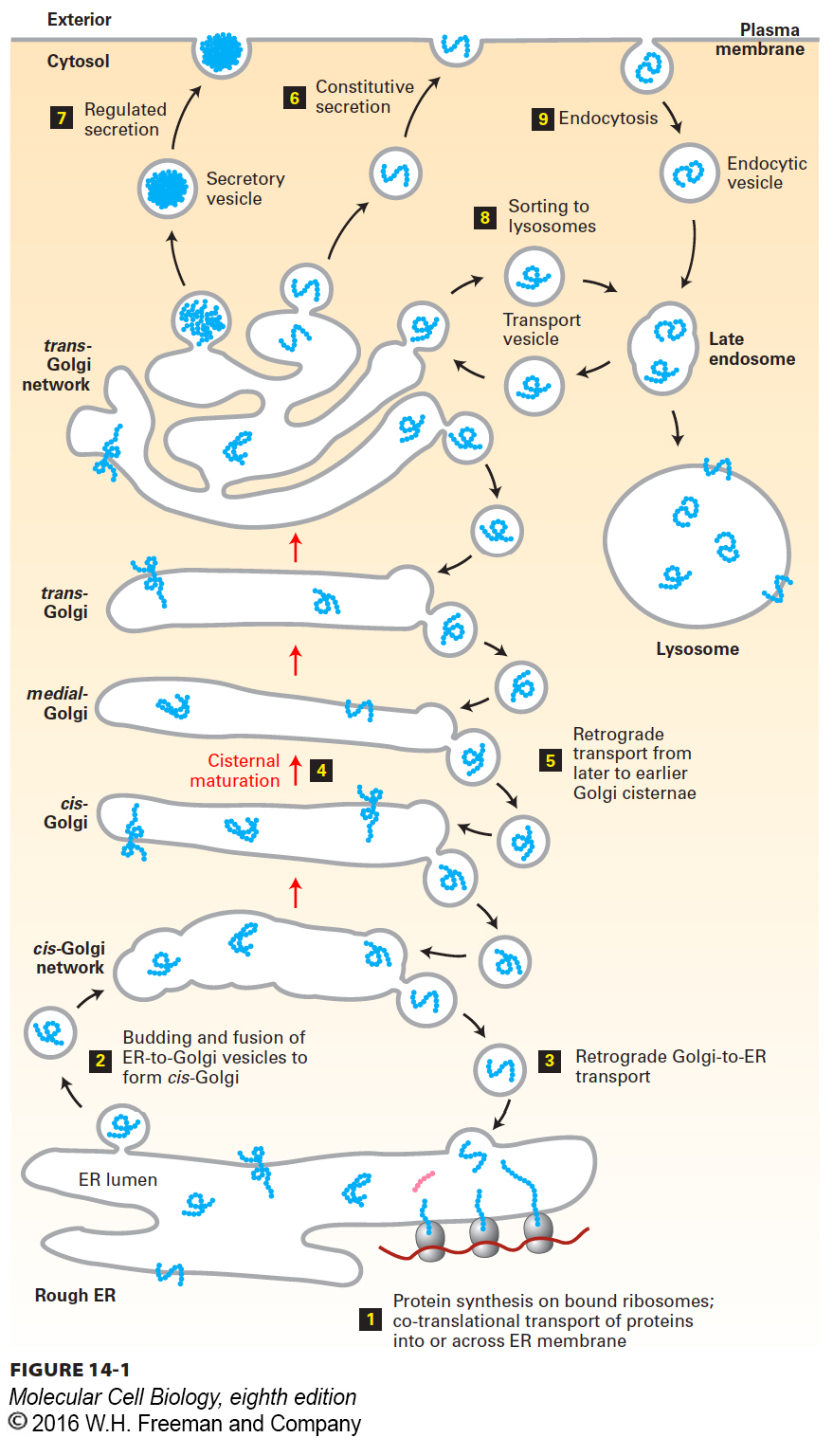
Reduced to its simplest elements, the secretory pathway operates in two stages. The first stage takes place in the rough endoplasmic reticulum (ER) (Figure 14-1, step 1). As described in Chapter 13, newly synthesized soluble and membrane proteins are translocated into the ER, where they fold into their proper conformation and receive covalent modifications such as N-linked and O-linked carbohydrates and disulfide bonds. Once newly synthesized proteins are properly folded and have received their correct modifications in the ER lumen, they progress to the second stage of the secretory pathway: transport to and through the Golgi complex.

The second stage of the secretory pathway can be summarized as follows. In the ER, cargo proteins are packaged into anterograde (forward-moving) transport vesicles (Figure 14-1, step 2). These vesicles fuse with one another to form a flattened membrane-bounded compartment known as the cis-Golgi network or cis-Golgi cisterna (a “cistern” is a container for holding water or other liquid). Certain proteins, mainly proteins that function in the ER, can be retrieved from the cis-Golgi cisterna and returned to the ER via a different set of retrograde (backward-moving) transport vesicles (step 3). In a manner reminiscent of an assembly line, the new cis-Golgi cisterna, with its cargo of proteins, physically moves from the cis position (nearest the ER) to the trans position (farthest from the ER), successively becoming first a medial-Golgi cisterna and then a trans-Golgi cisterna (step 4). This process, known as cisternal maturation, primarily involves retrograde transport vesicles (step 5), which retrieve enzymes and other Golgi-resident proteins from later to earlier Golgi cisternae, thereby “maturing” the cis-Golgi cisternae to medial-Golgi cisternae, and medial-Golgi cisternae to trans-Golgi cisternae. As secretory proteins move through the Golgi, their linked carbohydrates may be further modified by specific glycosyl transferases that are housed in the different Golgi compartments.

Proteins in the secretory pathway are eventually delivered to a complex network of membranes and vesicles termed the trans-Golgi network. The trans-Golgi network is a major branch point in the secretory pathway. It is at this stage that proteins are loaded into different kinds of vesicles and thereby trafficked to different destinations. Depending on which kind of vesicle the protein is loaded into, it will be transported to the plasma membrane and secreted immediately, stored for later release, or shipped to the lysosome (steps 68). The process by which a vesicle moves to and fuses with the plasma membrane and releases its contents is known as exocytosis. In all cell types, at least some proteins are secreted continuously (a process commonly called constitutive secretion), while others are stored inside the cell until a signal for exocytosis causes them to be released (regulated secretion). Secretory proteins destined for lysosomes are first transported by vesicles from the trans-Golgi network to a compartment usually called the late endosome; the proteins are then transferred to the lysosome by direct fusion of the late endosome with the lysosomal membrane.

Endocytosis is related mechanistically to the secretory pathway. In the endocytic pathway, vesicles bud inward from the plasma membrane, bringing membrane proteins and their bound ligands into the cell (see Figure 14-1, right). After being internalized by endocytosis, some proteins are transported to lysosomes via the late endosome, whereas others are recycled back to the cell surface.

633

In this chapter, we first discuss the experimental techniques that have contributed to our knowledge of the secretory pathway and endocytosis. Then we focus on the general mechanisms of membrane budding and fusion. We will see that although different kinds of transport vesicles use distinct sets of proteins for their formation and fusion, all vesicles use the same general mechanism for budding, selection of particular sets of cargo molecules, and fusion with the appropriate target membrane. In the remaining sections of the chapter, we discuss both the early and late stages of the secretory pathway, including how specificity of targeting to different destinations is achieved, and conclude with a discussion of how proteins are transported to the lysosome by the endocytic pathway.

634