Primary Cell Cultures and Cell Strains Have a Finite Life Span
Primary cells are cells isolated directly from tissues. Normal animal tissues (e.g., skin, kidney, liver) or whole embryos are commonly used to establish primary cell cultures. To prepare individual tissue cells for a primary culture, the cell-cell and cell-matrix interactions must be broken. To do so, tissue fragments are treated with a combination of a protease (e.g., trypsin, the collagen-hydrolyzing enzyme collagenase, or both) and a divalent cation chelator (e.g., EDTA) that depletes the medium of free calcium (Ca2+). Many CAMs require calcium and are thus inactivated when calcium is removed; other CAMs that are not calcium dependent need to be cleaved by a protease for the cells to separate. The released cells are then placed in a nutrient-rich, serum-supplemented medium in dishes, where they can adhere to the surface and to one another. The same protease-chelator solution is used to remove adherent cells from a culture dish for biochemical studies or subculturing (transfer to another dish).
Fibroblasts are the predominant cells in connective tissue and normally produce extracellular-matrix components such as collagen that bind to CAMs, thereby anchoring cells to a surface. In culture, fibroblasts usually divide more rapidly than other cells from a tissue, eventually becoming the predominant cell type in a primary culture unless special precautions are taken to remove them when isolating other types of cells.
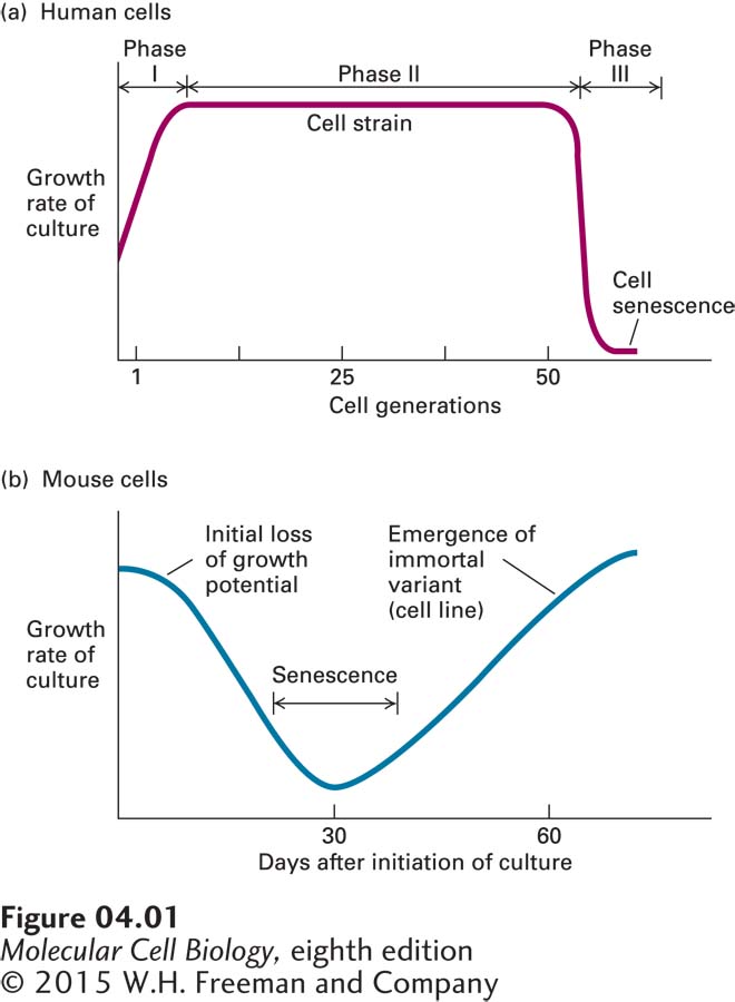
When cells removed from an embryo or an adult animal are cultured, most of the adherent cells will divide a finite number of times and then cease growing (a phenomenon called cell senescence). For instance, human fetal fibroblasts divide about 50 times before they cease growth (Figure 4-1a). Starting with 106 cells, 50 doublings has the potential to produce 106 × 250, or more than 1020, cells, whose weight would be equivalent to that of about a thousand people. Normally, only a very small fraction of these cells are used in any one experiment. Thus, even though its lifetime is limited, a single culture, if carefully maintained, can be studied through many cell generations. Such a lineage of cells originating from one initial primary culture is called a cell strain.
FIGURE 4-1Stages in the establishment of a cell culture. (a) When cells isolated from human tissue are initially cultured, some cells die and others (mainly fibroblasts) start to grow; overall, the growth rate increases (phase I). If the remaining cells are harvested, diluted, and replated into dishes again and again, the cell strain continues to divide at a constant rate for about 50 cell generations (phase II), after which the growth rate falls rapidly. In the ensuing period (phase III), all the cells in the culture stop growing (senescence). (b) In a culture prepared from mouse or other rodent cells, initial cell death (not shown) is coupled with the emergence of healthy, growing cells. As these dividing cells are diluted and allowed to continue growth, they soon begin to lose growth potential, and most stop growing (i.e., the culture goes into senescence). Very rare cells undergo oncogenic mutations that allow them to survive and continue dividing until their progeny overgrow the culture. These cells constitute a cell line, which will grow indefinitely if it is appropriately diluted and fed with nutrients. Such cells are said to be immortal.
One important exception to the finite life span of normal cells is the embryonic stem cell, which, as its name implies, is derived from an embryo and will divide and give rise to all tissues during development. As we discuss in Chapter 21, embryonic stems cells can be cultured indefinitely under the appropriate conditions.
Page 132
Research with cell strains is simplified by our ability to freeze them and successfully thaw them at a later time for experimental analysis. Cell strains can be frozen in a state of suspended animation and stored for extended periods at liquid nitrogen temperature, provided that a preservative that prevents the formation of damaging ice crystals is used. Although not all cells survive thawing, many do survive and resume growth.