In addition to serving as research models for studies of cell function, cultured cells can be converted into “factories” for producing specific proteins. For example, special cultured cells can be used to generate monoclonal antibodies, which are experimental tools widely used in many aspects of cell biological research. They are also used for diagnostic and therapeutic purposes in medicine, as we discuss in later chapters.
To understand the challenge of generating monoclonal antibodies, we must briefly review how mammals produce antibodies; more detail is provided in Chapter 23. Recall that antibodies are proteins secreted by white blood cells that bind with high affinity to their antigen (see Figure 3-19). Each normal antibody-producing B lymphocyte in a mammal is capable of producing a single type of antibody that can bind to a particular determinant, or epitope, on an antigen molecule. An epitope is generally a small region on the antigen, consisting, for example, of just a few amino acids. If an animal is injected with an antigen, the B lymphocytes that make antibodies recognizing that antigen are stimulated to grow and secrete those antibodies. Each antigen-activated B lymphocyte forms a clone of cells in the spleen or lymph nodes, with each cell producing the identical antibody—that is, a monoclonal antibody. Because most natural antigens contain multiple epitopes, exposure of an animal to an antigen usually stimulates the formation of multiple B-lymphocyte clones, each producing a different antibody. The resulting mixture of antibodies from the many B-lymphocyte clones that recognize different epitopes on the antigen is said to be polyclonal. Such polyclonal antibodies circulate in the blood and can be isolated as a group.
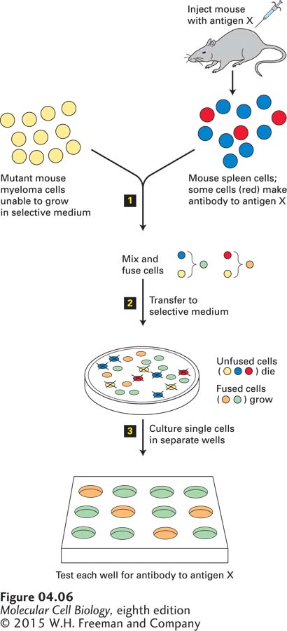
FIGURE 4-6Use of cell fusion and selection to obtain hybridomas producing a monoclonal antibody to a specific protein. Step 1: Immortal myeloma cells that cannot synthesize purines under special conditions because they lack thymidine kinase are fused with normal antibody-producing spleen cells from an animal that was immunized with antigen X. Step 2: When cultured in a special selective medium, unfused and self-fused cells do not grow: the myeloma cells do not grow because the selective medium does not contain purines, and the spleen cells do not grow because they have a limited life span in culture. Thus only fused cells formed from a myeloma cell and a spleen cell survive in the selective medium, proliferating into clones called hybridomas. Each hybridoma produces a single antibody. Step 3: Testing of individual clones identifies those that recognize antigen X. After a hybridoma that produces a desired antibody has been identified, the clone can be cultured to yield large amounts of that antibody.
Although polyclonal antibodies are very useful, monoclonal antibodies are more suitable for the many types of experiments and medical applications in which we need a reagent that binds to just one site on a protein; for example, one that competes with a ligand on a cell-surface receptor. Unfortunately, the biochemical purification of any one type of monoclonal antibody from blood is not feasible for two main reasons: the concentration of any given antibody is quite low, and all antibodies have the same basic molecular architecture (see Figure 3-19).
To produce and then purify monoclonal antibodies, one first needs to be able to grow the appropriate B-lymphocyte clone. However, primary cultures of normal B lymphocytes are of limited usefulness for the production of monoclonal antibodies because they have a limited life span. Thus the first step in producing a monoclonal antibody is to generate immortal antibody-producing cells (Figure 4-6). Immortality is achieved by fusing normal B lymphocytes from an immunized animal with transformed, immortal lymphocytes called myeloma cells that themselves do not synthesize antibodies (see Figure 3-19). Treatment with certain viral glycoproteins or the chemical polyethylene glycol encourages the plasma membranes of two cells to fuse, allowing their cytosols and organelles to intermingle. Some of the fused cells undergo division, and their nuclei eventually coalesce, producing viable hybrid cells with a single nucleus that contains chromosomes from both parent cells. The fusion of two cells that are genetically different can yield a hybrid cell with novel characteristics. For instance, the fusion of a myeloma cell with a normal antibody-producing cell from a rat or mouse spleen yields a hybrid that proliferates into a clone called a hybridoma. Like myeloma cells, hybridoma cells grow rapidly and are immortal. Each hybridoma produces the monoclonal antibody encoded by its B-lymphocyte parent.
The second step in this procedure for producing a monoclonal antibody is to separate, or select, the hybridoma cells from the unfused parent cells and the cells fused with another of the same type. This selection is usually performed by incubating the mixture of cells in a special culture medium, called a selection medium, that permits the growth of only the hybridoma cells because of their novel characteristics. The myeloma cells used for the fusion carry a mutation that blocks a metabolic pathway, so a selection medium can be used that is lethal to them and not to their B-lymphocyte fusion partners that do not have the mutation. In the immortal hybrid cells, the functional gene from the lymphocyte can supply the missing gene product. The lymphocytes used in the fusion are not immortal and will not be able to grow in the selection medium either. Thus the hybridoma cells will be the only ones able to proliferate rapidly in the selection medium and so can be readily isolated from the initial mixture of cells. Finally, each selected hybridoma clone is tested for the production of the desired antibody; any clone producing that antibody is then grown in large cultures, from which a substantial quantity of pure monoclonal antibody can be obtained.
Page 136
Monoclonal antibodies have become very valuable as specific research tools. They are commonly employed in affinity chromatography to isolate and purify proteins from complex mixtures (see Figure 3-38c). As we discuss later in this chapter, they can also be employed in immunofluorescence microscopy to bind to and so locate a particular protein within cells. They can also be used to identify specific proteins in cell fractions with the use of immunoblotting (see Figure 3-39). Monoclonal antibodies have become important diagnostic and therapeutic tools in medicine as well; for example, monoclonal antibodies that bind to and inactivate toxins secreted by bacterial pathogens are used to treat diseases. Other monoclonal antibodies are specific for cell-surface proteins expressed by certain types of tumor cells. Several of these anti-tumor antibodies are widely used in cancer therapy, including a monoclonal antibody against a mutant form of the Her2 receptor that is overexpressed in some breast cancers.