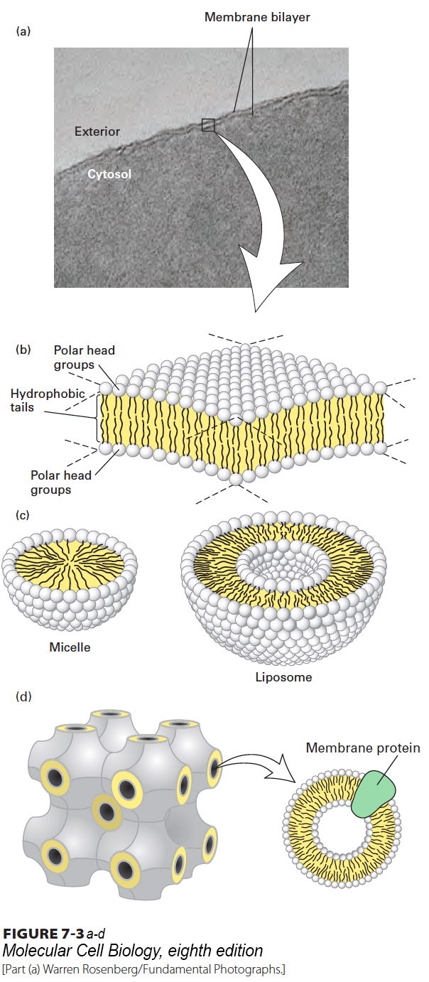
The amphipathic nature of phospholipids, which governs their interactions, is critical to the structure of biomembranes. When a suspension of phospholipids is mechanically dispersed in an aqueous solution, the phospholipids aggregate into one of three structures: spherical micelles or liposomes or sheet-like phospholipid bilayers that are two molecules thick (Figure 7-3). The type of structure formed by pure phospholipids or a mixture of phospholipids in the absence of added proteins depends on several factors, including the length of the fatty acyl chains in the hydrophobic tails, their degree of saturation (i.e., the number of C−C and C=C bonds), and temperature. In all three structures, the hydrophobic effect (see Chapter 2) causes the fatty acyl chains to aggregate and exclude water molecules from the “core” of the structure. Micelles are rarely formed from natural phospholipids, whose fatty acyl chains are generally too bulky to fit into the interior of a micelle. Micelles are formed, however, if one of the two fatty acyl chains that make up the tail of a phospholipid is removed by hydrolysis to form a lysophospholipid, as occurs upon treatment with the enzyme phospholipase. In aqueous solutions, common detergents and soaps form micelles that behave like the balls in tiny ball bearings, thus giving soap solutions their slippery feel and lubricating properties.
FIGURE 7-3The bilayer structure of biomembranes. (a) Electron micrograph of a thin section through an erythrocyte membrane stained with osmium tetroxide. The characteristic “railroad track” appearance of the membrane indicates the presence of two polar layers, consistent with the bilayer structure of phospholipid membranes. (b) Schematic interpretation of the phospholipid bilayer, in which polar groups face outward to shield the hydrophobic fatty acyl tails from water. The hydrophobic effect and van der Waals interactions between the fatty acyl tails drive the assembly of the bilayer (see Chapter 2). (c) Cross-sectional views of two other structures formed by dispersal of phospholipids in water. A spherical micelle has a hydrophobic interior composed entirely of fatty acyl chains; a spherical liposome consists of a phospholipid bilayer surrounding an aqueous center. (d) Under certain circumstances, lipids can assume yet other forms of organization. Shown here is the cubic phase of lipids, a highly regular recurring structure that has helped the formation of crystals of membrane proteins that were otherwise difficult to crystallize.
Phospholipid mixtures of the composition present in cells spontaneously form a symmetric phospholipid bilayer. Each phospholipid layer in this lamellar structure is called a leaflet. The hydrophobic fatty acyl chains in each leaflet minimize their contact with water by aligning themselves tightly together in the center of the bilayer, forming a hydrophobic core that is about 3–4 nm thick (see Figure 7-3b). The close packing of these nonpolar tails is stabilized by van der Waals interactions between the hydrocarbon chains. Ionic and hydrogen bonds stabilize the interactions of the polar head groups with one another and with water. Electron microscopy of thin sections of cells stained with osmium tetroxide, which binds strongly to the polar head groups of phospholipids, shows the bilayer structure (see Figure 7-3a). A cross section of a single membrane stained with osmium tetroxide looks like a railroad track: two thin dark lines (the stained head group complexes) with a uniform light space of about 2 nm between them (the hydrophobic tails).
A phospholipid bilayer can be of almost unlimited size—from micrometers (µm) to millimeters (mm) in length or width—and can contain tens of millions of phospholipid molecules. The phospholipid bilayer is the basic structural unit of nearly all biological membranes. Its hydrophobic core prevents most water-soluble substances from crossing from one side of the membrane to the other. Although biomembranes contain other molecules (e.g., cholesterol, glycolipids, proteins), it is the phospholipid bilayer that separates two aqueous solutions and acts as a permeability barrier. The lipid bilayer thus defines cellular compartments and allows a separation of the cell’s interior from the outside world.
The three structures mentioned above are not the only forms that lipids can assume in an aqueous environment. Unusual configurations of lipids have been instrumental in enforcing order on otherwise difficult-to-crystallize membrane proteins, including G protein–coupled receptors, enabling crystallographic analysis of membrane proteins in a true lipid environment (Figure 7-3d).