Chapter 37 Summary
Core Concepts Summary
37.1 Muscles are biological motors composed of actin and myosin that generate force and produce movement for support, locomotion, and control of internal physiological functions.
There are two main types of muscle: striated (skeletal and cardiac) and smooth. page 787
Skeletal and cardiac muscle fibers appear striated when viewed under the microscope because of the regular spacing of sarcomeres along their length. page 787
Smooth muscle fibers, which regulate airflow for breathing, blood flow through arteries, and the passage of food through the gut, lack regular sarcomere organization and appear smooth when viewed under the microscope. page 787
Skeletal muscle fibers are long thin cells composed of parallel sets of myofibrils built up from smaller parallel arrays of actin and myosin filaments. page 787
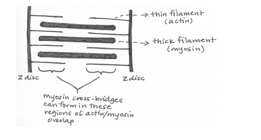
The sarcomere is the basic contractile unit of a skeletal muscle. Sarcomeres are arranged in series along the length of a myofibril. page 788
Muscles change length and produce force by the formation of actin–
Muscles are stimulated by motor neurons at the fiber’s motor endplate, leading to depolarization of the muscle cell that triggers the release of Ca2+ ions from the sarcoplasmic reticulum. Ca2+ binds troponin, which moves tropomyosin off the myosin-
Excitation–
37.2 The force generated by muscle depends on muscle size, degree of actin–
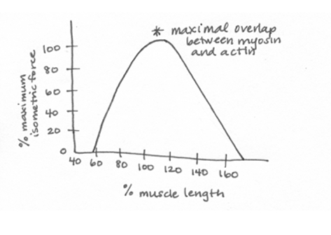
Muscles exert their greatest force when actin–
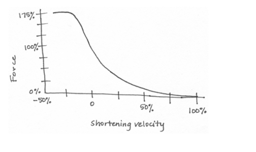
Muscles exert more force when they contract at slow velocities compared with when they contract at high velocities. page 793
Because muscles can transmit force only by pulling on the skeleton, they are arranged as antagonist pairs to produce reciprocal motions of a joint or limb. page 794
In vertebrates, a motor unit consists of a single motor neuron and the muscle fibers (cells) it innervates. page 795
Muscle force is increased by increasing the motor neuron firing rate and, in vertebrates, by increasing the number of motor units that are activated. page 796
Vertebrate muscles have two types of fiber: red slow-
37.3 Hydrostatic skeletons, exoskeletons, and endoskeletons provide animals with mechanical support and protection.
The rigid element of a hydrostatic skeleton is an incompressible fluid within a body cavity, used to support the body and change shape. page 797
Invertebrate exoskeletons form an external rigid support system, which protects the animal and limits water loss, but also limits growth and repair. page 798
Vertebrates have a bony endoskeleton that provides rigid support and protection of body organs, and that can grow and be repaired. page 799
The vertebrate endoskeleton is organized into axial (central) and appendicular (limb) components. page 800
Vertebrate endoskeletons consist of bone and cartilage. page 800
Bone is a composite tissue that consists of calcium phosphate mineral and type I collagen and that is rigid and hard to break. page 800
Cartilage is a fluid-
37.4 Vertebrate endoskeletons allow for growth and repair and transmit muscle forces across joints.
Vertebrate bones develop by one of two processes: either directly or by way of a cartilage model. page 801
Cartilage forms much of the embryonic skeleton and remains as growth plates within the bone to provide rapid growth after birth, as well as forming the bone’s joint (articular) surfaces. page 801
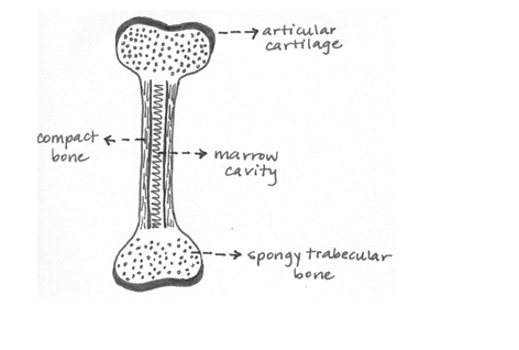
Bone has two basic structures: Compact bone forms the solid walls of a bone’s shaft, and spongy bone forms a mesh that supports the cartilage at the bone’s ends. page 801
Bone formation by osteoblasts and removal by osteoclasts is a continuing process of growth, shape change, and repair. page 802
The shape of a bone’s joint surfaces largely determines the range of motion and stability of a joint. page 802
Muscles produce joint movements by transmitting forces by means of rigid skeletal levers. page 802
Self-Assessment
Diagram a sarcomere, showing the basic organization of thin (actin) and thick (myosin) filaments. Indicate on your diagram the regions where myosin cross-
bridges form with actin. Self-Assessment 1 Answer
Describe the sequence of events that occurs when an action potential arrives at a muscle fiber’s motor endplate, causing the muscle to be depolarized.
Self-Assessment 2 Answer
Depolarization at the motor endplate of a muscle fiber is propagated from the cell surface to the sarcoplasmic reticulum (SR) via the T-
tubule system. Depolarization of the SR causes release of Ca2+ ions from the SR into the cytoplasm. The Ca2+ ions bind to troponin and cause a conformational change in troponin that causes tropomyosin to move. The movement of tropomyosin exposes the myosin- binding sites on actin, allowing for the formation of myosin- actin cross- bridges and muscle contraction. Describe the sequence of events involved in myosin cross-
bridge attachment and detachment. Relate these events to how muscles generate force and shorten when they are stimulated to contract, identifying the role that ATP plays. Self-Assessment 3 Answer
With ADP and inorganic phosphate (Pi) bound to the myosin head, the myosin is able to rotate toward a binding site located along the actin filament toward the Z-
disc at the end of the sarcomere. The myosin head binds to an available actin binding site, forming a cross- bridge. ADP and Pi are then split from the myosin cross- bridge head, causing the head to pivot toward the sarcomere midline, producing force and moving the actin filament relative to the myosin filament, leading to sarcomere shortening. This is referred to as the power stroke. The myosin head then binds a new ATP, allowing it to release from the actin binding site. With the subsequent splitting of ATP into ADP and Pi, the myosin head is again able to rotate, beginning the next cross- bridge cycle. Draw a graph of the isometric force–
length relationship of striated muscle, indicating where maximal overlap between actin and myosin filaments occurs. Self-Assessment 4 Answer
Draw a graph of the force–
shortening velocity relationship of striated muscle. Self-Assessment 5 Answer
Compare the force produced by a muscle over time when it is stimulated by a single twitch stimulus with the force produced by multiple stimuli at low frequency, and then with the force produced when the stimulation frequency is increased.
Self-Assessment 6 Answer
A single twitch stimulus will result in a certain amount of force based on the number of muscle fibers in the motor unit. If the number of stimuli increase but the frequency is low, the muscle fully relaxes between stimuli. In this case, the force produced by each stimulus will be equal to that of a single twitch stimulus. When the frequency of the stimuli is increased further so that the muscle does not have time to relax between stimuli, the forces produced by the individual stimuli are summed together to generate a greater force than a single twitch stimulus can produce. At a sufficiently high frequency, a sustained contraction of the muscle, known as tetanus, occurs.
Name the two basic structural elements common to all animal musculoskeletal systems.
Self-Assessment 7 Answer
All animal musculoskeletal systems consist of rigid elements (such as cartilage, bones, or fluid-
filled cavities) and groups of muscles that work as antagonists. Compare and contrast three features of an exoskeleton and an endoskeleton.
Self-Assessment 8 Answer
(1) While both endoskeletons and exoskeletons provide support and protection for an animal, exoskeletons cover the outside of an animal and provide external protection from harm and dessication, whereas endoskeletons are internal and provide protection to vital organs but not the animal’s exterior. (2) Both types of skeleton are made from composite materials, which makes them strong and flexible. However, endoskeletons, like those found in vertebrates, are first formed as cartilage and then mineralized to form hardened bone, whereas exoskeletons are formed by the deposition of mineralized proteins at the outer edge of the growing skeleton. (3) Endoskeletons allow for greater range of motion because they have joints between adjacent bones, whereas organisms with exoskeletons are more restricted in their movements since they usually only have joints between different body segments. (4) Exoskeletons limit growth because organisms must undergo molting and regeneration of their skeleton as they increase in size, and very large exoskeletons are especially prone to damage because they are so thin. In contrast, bone length in endoskeletons can increase over time to support growth of the organism during development and throughout its lifetime. (5) Damage to both types of skeleton can be repaired, but animals with exoskeletons must form an entirely new skeleton, whereas animals with endoskeletons can repair specific regions of damaged bone through the work of osteoblasts.
Identify the two primary components of bone tissue and explain how each of these contributes to a bone’s strength, stiffness, and resistance to fracture.
Self-Assessment 9 Answer
Bone tissue is composed of hydroxyapitite mineral and collagen protein. Hydroxyapitite makes the bone strong and rigid, and collagen protein allows the bone to absorb force and resist breaking.
Diagram a limb bone, such as the tibia, showing the regions of articular cartilage, spongy trabecular bone, compact bone tissue, and the marrow cavity.
Self-Assessment 10 Answer