2.2 Tools of Discovery and Older Brain Structures

In a jar on a display shelf in Cornell University’s psychology department resides the well-
You might answer that, without the living whir of electrochemical activity, there could be nothing of Titchener in his preserved brain. Consider, then, an experiment about which the inquisitive Titchener himself might have daydreamed. Imagine that just moments before his death, someone had removed Titchener’s brain and kept it alive by feeding it enriched blood. Would Titchener still be in there? Further imagine that someone then transplanted the still-
“I am a brain, Watson. The rest of me is a mere appendix.”
Sherlock Holmes, in Arthur Conan Doyle’s “The Adventure of the Mazarin Stone”
That we can imagine such questions illustrates how convinced we are that we live “somewhere north of the neck” (Fodor, 1999). And for good reason: The brain enables the mind—
The Tools of Discovery: Having Our Head Examined
2-
A century ago, scientists had no tools high powered yet gentle enough to explore the living human brain. Early case studies helped localize some brain functions. Damage to one side of the brain often caused numbness or paralysis on the body’s opposite side, suggesting that the body’s right side is wired to the brain’s left side, and vice versa. Damage to the back of the brain disrupted vision, and to the left-
lesion [LEE-zhuhn] tissue destruction. A brain lesion is a naturally or experimentally caused destruction of brain tissue.
Now, within a lifetime, a new generation of neural cartographers is probing and mapping the known universe’s most amazing organ. Scientists can selectively lesion (destroy) tiny clusters of brain cells, leaving the surrounding tissue unharmed. In the laboratory, such studies have revealed, for example, that damage to one area of the hypothalamus in a rat’s brain reduces eating, to the point of starvation, whereas damage in another area produces overeating.
Today’s neuroscientists can also stimulate various brain parts—
67
Right now, your mental activity is emitting telltale electrical, metabolic, and magnetic signals that would enable neuroscientists to observe your brain at work. Electrical activity in your brain’s billions of neurons sweeps in regular waves across its surface. An electroencephalogram (EEG) is an amplified readout of such waves. Researchers record the brain waves through a shower-
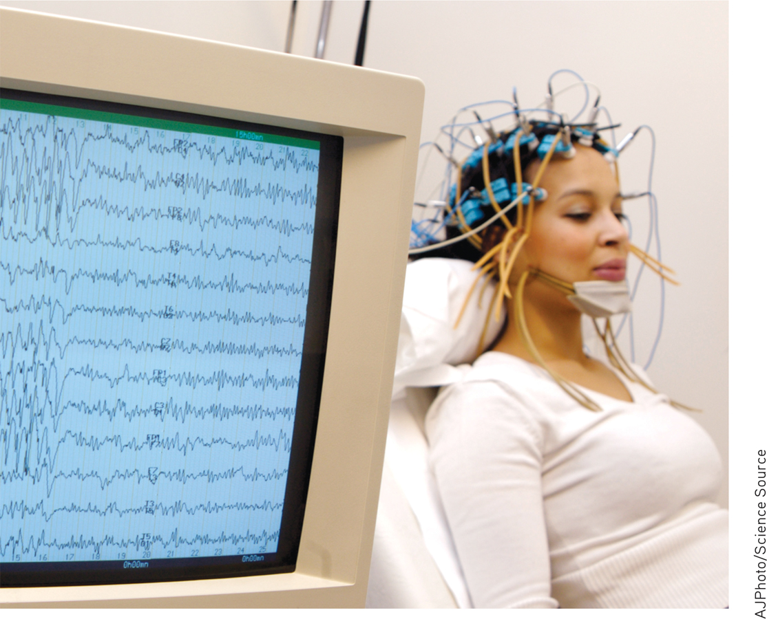
Brain hacking An electroencephalograph provides amplified tracings of waves of electrical activity in the brain.
electroencephalogram (EEG) an amplified recording of the waves of electrical activity sweeping across the brain’s surface. These waves are measured by electrodes placed on the scalp.
“You must look into people, as well as at them,” advised Lord Chesterfield in a 1746 letter to his son. Unlike EEGs, newer neuroimaging techniques give us that Superman-
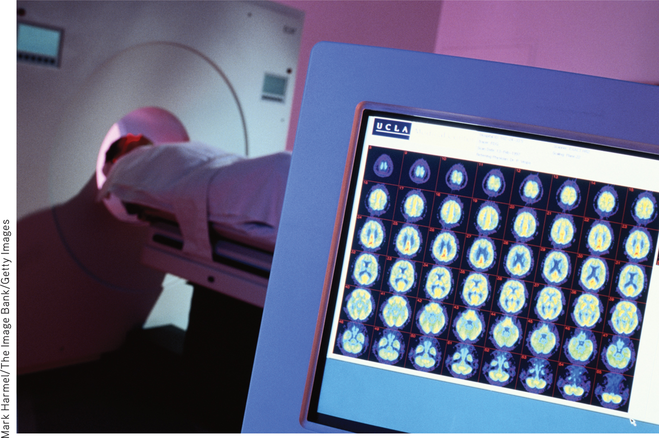
The PET scan To obtain a PET scan, researchers inject volunteers with a low and harmless dose of a short-
PET (positron emission tomography) scan a visual display of brain activity that detects where a radioactive form of glucose goes while the brain performs a given task.
In MRI (magnetic resonance imaging) brain scans, the person’s head is put in a strong magnetic field, which aligns the spinning atoms of brain molecules. Then, a radio-
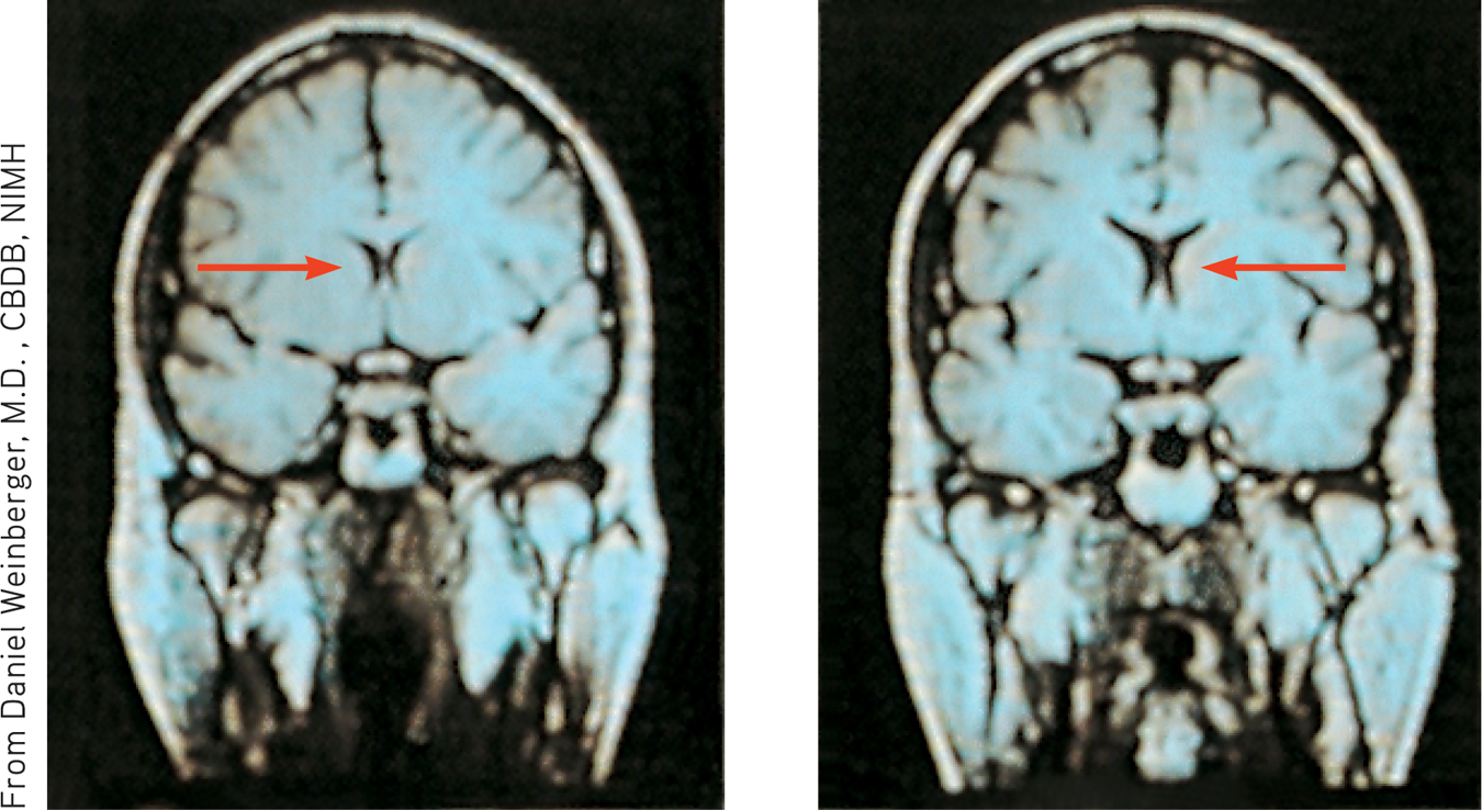
MRI scan of a healthy individual (left) and a person with schizophrenia (right) Note the enlarged ventricle, the fluid-
MRI (magnetic resonance imaging) a technique that uses magnetic fields and radio waves to produce computer-generated images of soft tissue. MRI scans show brain anatomy.
68
fMRI (functional MRI) a technique for revealing bloodflow and, therefore, brain activity by comparing successive MRI scans. fMRI scans show brain function as well as structure.
A special application of MRI—
Such snapshots of the brain’s changing activity are providing new insights into how the brain divides its labor. A mountain of recent fMRI studies suggests which brain areas are most active when people feel pain or rejection, listen to angry voices, think about scary things, feel happy, or become sexually excited. The technology enables a very crude sort of mind reading. One neuroscience team scanned 129 people’s brains as they did eight different mental tasks (such as reading, gambling, or rhyming). Later, they were able, with 80 percent accuracy, to predict which of these mental activities a person was doing (Poldrack et al., 2009). Other studies have explored brain activity associated with religious experience, though without settling the question of whether the brain is producing or perceiving God (Fingelkurts & Fingelkurts, 2009; Inzlicht et al., 2009; Kapogiannis et al., 2009).
You’ve seen the pictures—
***
Today’s techniques for peering into the thinking, feeling brain are doing for psychology what the microscope did for biology and the telescope did for astronomy. From them we have learned more about the brain in the last 30 years than in the previous 30,000. And the next decade will reveal much more, as each year massive funding goes into brain research. Europe’s Human Brain Project promises $1 billion for brain computer modeling and the $40 million Human Connectome Project (2013; Gorman, 2014) seeks “neural pathways [that] will reveal much about what makes us uniquely human and what makes every person different from all others.” A new super-
To be learning about the neurosciences now is like studying world geography while Magellan was exploring the seas. The whole brain mapping effort now underway has been likened to last century’s Apollo program that landed humans on the Moon, and to the Human Genome Project’s mapping our DNA. This truly is the golden age of brain science.
RETRIEVAL PRACTICE
- Match the scanning technique with the correct description.
TechniqueDescription
1. fMRI scan a. tracks radioactive glucose
to reveal brain activity.
2. PET scan b. tracks successive images of
brain tissue to show brain
function.
3. MRI scan c. uses magnetic fields and
radio waves to show
brain anatomy.
1. b, 2. a, 3. c
69
Older Brain Structures

2-
An animal’s capacities come from its brain structures. In primitive animals, such as sharks, a not-
This increasing complexity arises from new brain systems built on top of the old, much as the Earth’s landscape covers the old with the new. Digging down, one discovers the fossil remnants of the past—
 For an introductory 12.5-minute overview of the brain, visit LaunchPad’s Video: The Central Nervous System—Spotlight on the Brain.
The Brainstem
brainstem the oldest part and central core of the brain, beginning where the spinal cord swells as it enters the skull; the brainstem is responsible for automatic survival functions.
The brain’s oldest and innermost region is the brainstem. It begins where the spinal cord swells slightly after entering the skull. This slight swelling is the medulla (FIGURE 2.15). Here lie the controls for your heartbeat and breathing. As some brain-
medulla [muh-DUL-uh] the base of the brainstem; controls heartbeat and breathing.
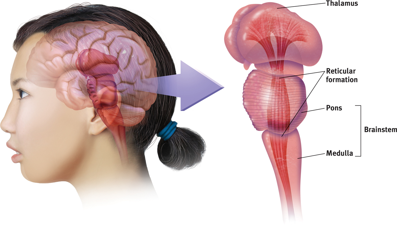
The brainstem and thalamus The brainstem, including the pons and medulla, is an extension of the spinal cord. The thalamus is attached to the top of the brainstem. The reticular formation passes through both structures.
If a cat’s brainstem is severed from the rest of the brain above it, the animal will still breathe and live—
The brainstem is a crossover point, where most nerves to and from each side of the brain connect with the body’s opposite side (FIGURE 2.16 below). This peculiar cross-
RETRIEVAL PRACTICE
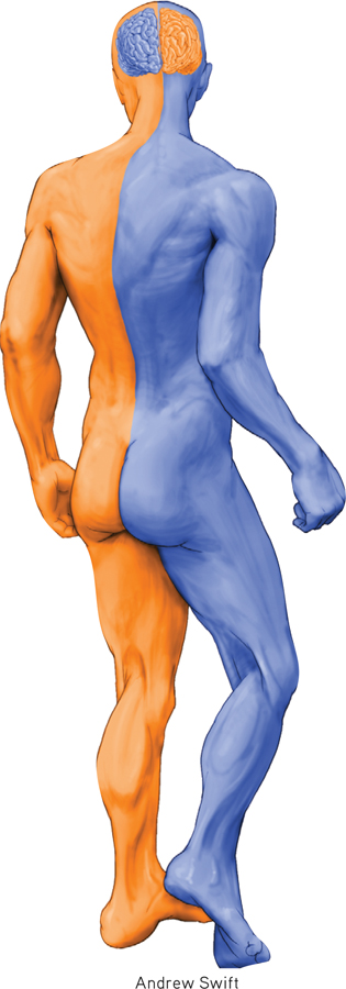
The body’s wiring
- Nerves from the left side of the brain are mostly linked to the ______________ side of the body, and vice versa.
right
70
The Thalamus
thalamus [THAL-uh-muss] the brain’s sensory control center, located on top of the brainstem; it directs messages to the sensory receiving areas in the cortex and transmits replies to the cerebellum and medulla.
Sitting atop the brainstem is the thalamus, a pair of egg-
The Reticular Formation
reticular formation a nerve network that travels through the brainstem into the thalamus and plays an important role in controlling arousal.
Inside the brainstem, between your ears, lies the reticular (“net-
In 1949, Giuseppe Moruzzi and Horace Magoun discovered that electrically stimulating a sleeping cat’s reticular formation almost instantly produced an awake, alert animal. When Magoun severed a cat’s reticular formation without damaging nearby sensory pathways, the effect was equally dramatic: The cat lapsed into a coma from which it never awakened. The conclusion? The reticular formation enables arousal.
The Cerebellum
Extending from the rear of the brainstem is the baseball-
***
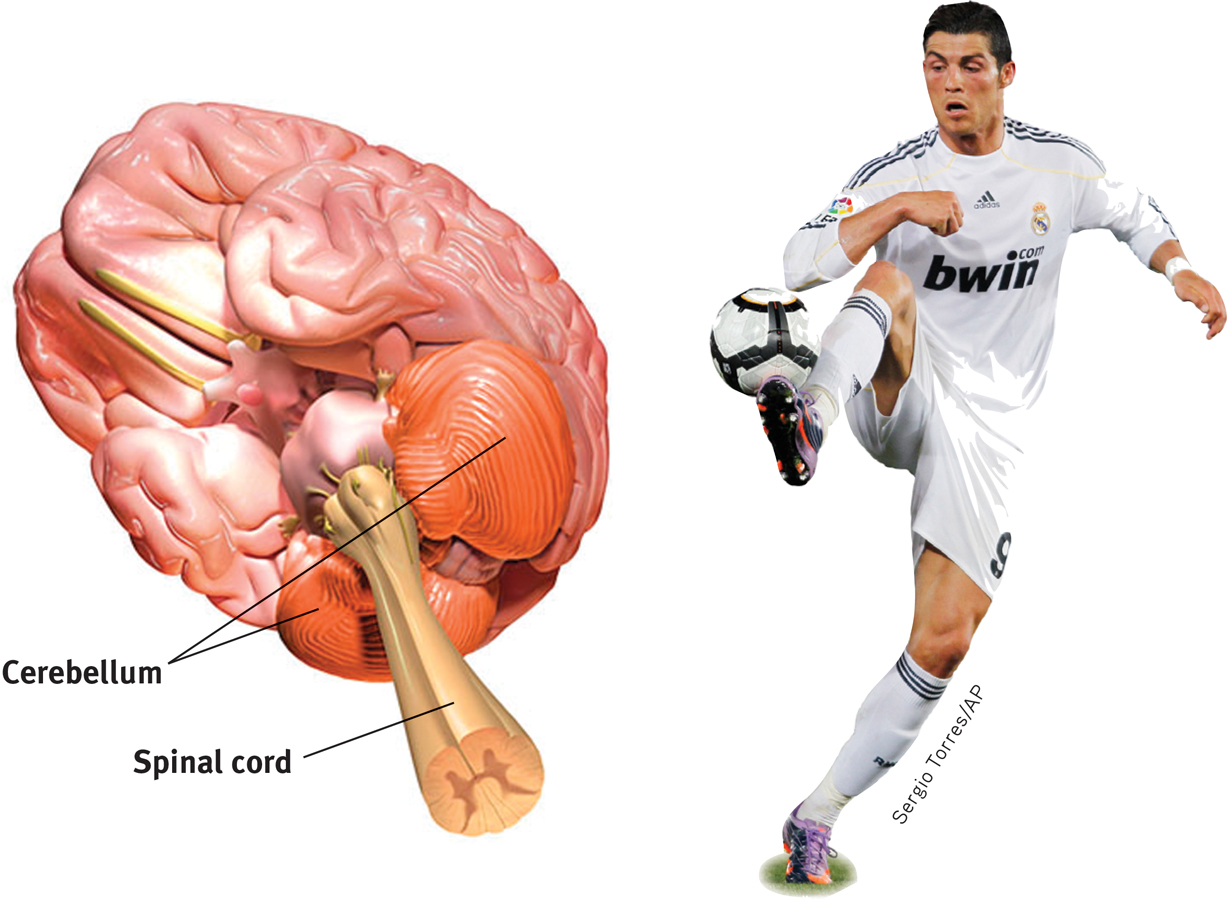
The brain’s organ of agility Hanging at the back of the brain, the cerebellum coordinates our voluntary movements.
cerebellum [sehr-uh-BELL-um] the “little brain” at the rear of the brainstem; functions include processing sensory input, coordinating movement output and balance, and enabling nonverbal learning and memory.
Note: These older brain functions all occur without any conscious effort. This illustrates another of our recurring themes: Our brain processes most information outside of our awareness. We are aware of the results of our brain’s labor (say, our current visual experience) but not of the how. Likewise, whether we are asleep or awake, our brainstem manages its life-
To review and check your understanding, visit LaunchPad’s Concept Practice: Lower Brain Structures.
RETRIEVAL PRACTICE
- In what brain region would damage be most likely to (1) disrupt your ability to skip rope? (2) disrupt your ability to hear and taste? (3) perhaps leave you in a coma? (4) cut off the very breath and heartbeat of life?
1. cerebellum, 2. thalamus, 3. reticular formation, 4. medulla
71
The Limbic System
2-
limbic system neural system (including the hippocampus, amygdala, and hypothalamus) located below the cerebral hemispheres; associated with emotions and drives.
We’ve considered the brain’s oldest parts, but we’ve not yet reached its newest and highest regions, the cerebral hemispheres (the two halves of the brain). Between the oldest and newest brain areas lies the limbic system (limbus means “border”). This system contains the amygdala, the hypothalamus, and the hippocampus (FIGURE 2.18). The hippocampus processes conscious, explicit memories. Animals or humans who lose their hippocampus to surgery or injury also lose their ability to form new memories of facts and events. Chapter 8 explains how our two-

The limbic system This neural system sits between the brain’s older parts and its cerebral hemispheres. The limbic system’s hypothalamus controls the nearby pituitary gland.
hippocampus a neural center located in the limbic system; helps process explicit memories for storage.
amygdala [uh-MIG-duh-la] two lima-bean-sized neural clusters in the limbic system; linked to emotion.
The Amygdala Research has linked the amygdala, two lima-

What then might happen if we electrically stimulated the amygdala of a placid domestic animal, such as a cat? Do so in one spot and the cat prepares to attack, hissing with its back arched, its pupils dilated, its hair on end. Move the electrode only slightly within the amygdala, cage the cat with a small mouse, and now it cowers in terror.
These and other experiments have confirmed the amygdala’s role in fear and rage. One study found math anxiety associated with hyperactivity in the right amygdala (Young et al., 2012). Other studies have shown people angry and happy faces: The amygdala activates in response to the angry ones (Mende-
RETRIEVAL PRACTICE
- Electrical stimulation of a cat’s amygdala provokes angry reactions. Which autonomic nervous system division is activated by such stimulation?
The sympathetic nervous system
hypothalamus [hi-po-THAL-uh-muss] a neural structure lying below (hypo) the thalamus; it directs several maintenance activities (eating, drinking, body temperature), helps govern the endocrine system via the pituitary gland, and is linked to emotion and reward.
The Hypothalamus Just below (hypo) the thalamus is the hypothalamus (FIGURE 2.19 below), an important link in the command chain governing bodily maintenance. Some neural clusters in the hypothalamus influence hunger; others regulate thirst, body temperature, and sexual behavior. Together, they help maintain a steady (homeostatic) internal state.
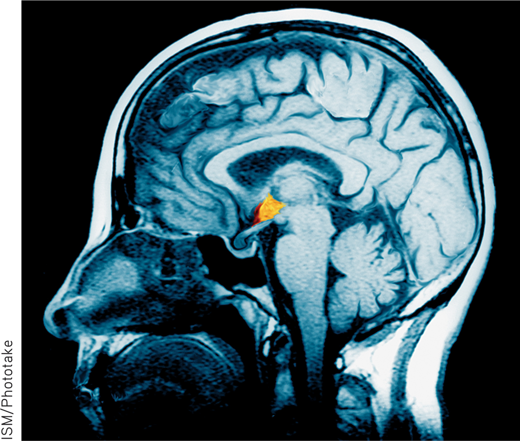
The hypothalamus This small but important structure, colored yellow/orange in this MRI-
As the hypothalamus monitors the state of your body, it tunes into your blood chemistry and any incoming orders from other brain parts. For example, picking up signals from your brain’s cerebral cortex that you are thinking about sex, your hypothalamus will secrete hormones. These hormones will in turn trigger the adjacent “master gland” of the endocrine system, your pituitary (see Figure 2.18), to influence your sex glands to release their hormones. These will intensify the thoughts of sex in your cerebral cortex. (Once again, we see the interplay between the nervous and endocrine systems: The brain influences the endocrine system, which in turn influences the brain.)
72
A remarkable discovery about the hypothalamus illustrates how progress in science often occurs—
In a meticulous series of experiments, Olds (1958) went on to locate other “pleasure centers,” as he called them. (What the rats actually experience only they know, and they aren’t telling. Rather than attribute human feelings to rats, today’s scientists refer to reward centers, not “pleasure centers.”) When allowed to press pedals to trigger their own stimulation, rats would sometimes do so more than 1000 times per hour. Moreover, they would even cross an electrified floor that a starving rat would not cross to reach food (FIGURE 2.20).
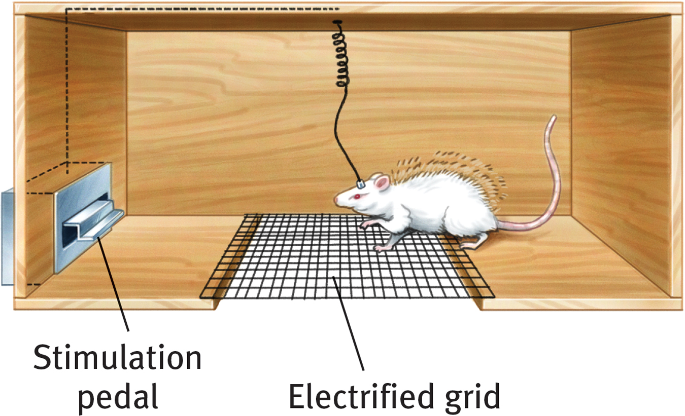
Rat with an implanted electrode With an electrode implanted in a reward center of its hypothalamus, the rat readily crosses an electrified grid, accepting the painful shocks, to press a pedal that sends electrical impulses to that center.
In other species, including dolphins and monkeys, researchers later discovered other limbic system reward centers, such as the nucleus accumbens in front of the hypothalamus. Animal research has also revealed both a general dopamine-
Researchers are experimenting with new ways of using brain stimulation to control animals’ actions in search-
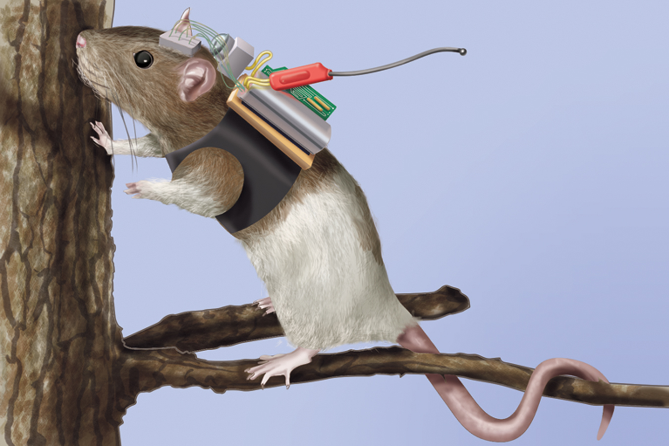
Ratbot on a pleasure cruise Researchers used a remote control brain stimulator to guide rats across a field and even up a tree.
Do humans have limbic centers for pleasure? To calm violent patients, one neurosurgeon implanted electrodes in such areas. Stimulated patients reported mild pleasure; unlike Olds’ rats, however, they were not driven to a frenzy (Deutsch, 1972; Hooper & Teresi, 1986). Moreover, newer research reveals that stimulating the brain’s “hedonic hotspots” (its reward circuits) produces more desire than pure enjoyment (Kringelbach & Berridge, 2012). Experiments have also revealed the effects of a dopamine-
73
“If you were designing a robot vehicle to walk into the future and survive, … you’d wire it up so that behavior that ensured the survival of the self or the species—like sex and eating—would be naturally reinforcing.”
Candace Pert (1986)
Some researchers believe that addictive disorders, such as substance use disorders and binge eating, may stem from malfunctions in natural brain systems for pleasure and well-
***
FIGURE 2.22 locates the brain areas we’ve discussed, as well as the cerebral cortex, our next topic.
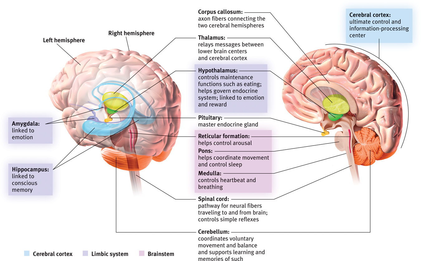
Brain structures and their functions
To review and assess your understanding, visit LaunchPad’s Concept Practice: The Limbic System.
RETRIEVAL PRACTICE
- What are the three key structures of the limbic system, and what functions do they serve?
(1) The amygdala is involved in aggression and fear responses. (2) The hypothalamus is involved in bodily maintenance, pleasurable rewards, and control of the hormonal systems. (3) The hippocampus processes conscious memory.
74
REVIEW: Tools of Discovery and Older Brain Structures

|
REVIEW | Tools of Discovery and Older Brain Structures |
LEARNING OBJECTIVES
RETRIEVAL PRACTICE Take a moment to answer each of these Learning Objective Questions (repeated here from within this section). Then click the 'show answer' button to check your answers. Research suggests that trying to answer these questions on your own will improve your long-
2-
Clinical observations and lesioning reveal the general effects of brain damage. Electrical, chemical, or magnetic stimulation can also reveal aspects of information processing in the brain. MRI scans show anatomy. EEG, PET, and fMRI (functional MRI) recordings reveal brain function.
2-
The brainstem, the oldest part of the brain, is responsible for automatic survival functions. Its components are the medulla (which controls heartbeat and breathing), the pons (which helps coordinate movements), and the reticular formation (which affects arousal).
The thalamus, sitting above the brainstem, acts as the brain’s sensory control center. The cerebellum, attached to the rear of the brainstem, coordinates muscle movement and balance and also helps process sensory information.
2-
The limbic system is linked to emotions, memory, and drives. Its neural centers include the hippocampus (which processes conscious memories); the amygdala (involved in responses of aggression and fear); and the hypothalamus (involved in various bodily maintenance functions, pleasurable rewards, and the control of the endocrine system). The hypothalamus controls the pituitary (the “master gland”) by stimulating it to trigger the release of hormones.
TERMS AND CONCEPTS TO REMEMBER
RETRIEVAL PRACTICE Match each of the terms on the left with its definition on the right. Click on the term first and then click on the matching definition. As you match them correctly they will move to the bottom of the activity.
Question
lesion [LEE-zhuhn] electroencephalogram (EEG) PET (positron emission tomography) scan MRI (magnetic resonance imaging) fMRI (functional MRI) brainstem medulla [muh-DUL-uh] thalamus [THAL-uh-muss] reticular formation cerebellum [sehr-uh-BELL-um] limbic system hippocampus amygdala [uh-MIG-duh-la] hypothalamus [hi-po-THAL-uh-muss] | a technique that uses magnetic fields and radio waves to produce computer-generated images of soft tissue. MRI scans show brain anatomy. tissue destruction. A brain lesion is a naturally or experimentally caused destruction of brain tissue. two lima-bean-sized neural clusters in the limbic system; linked to emotion. the brain’s sensory control center, located on top of the brainstem; it directs messages to the sensory receiving areas in the cortex and transmits replies to the cerebellum and medulla. neural system (including the hippocampus, amygdala, and hypothalamus) located below the cerebral hemispheres; associated with emotions and drives. the oldest part and central core of the brain, beginning where the spinal cord swells as it enters the skull; the brainstem is responsible for automatic survival functions. a nerve network that travels through the brainstem into the thalamus and plays an important role in controlling arousal. the “little brain” at the rear of the brainstem; functions include processing sensory input, coordinating movement output and balance, and enabling nonverbal learning and memory. a neural center located in the limbic system; helps process explicit memories for storage. a technique for revealing bloodflow and, therefore, brain activity by comparing successive MRI scans. fMRI scans show brain function as well as structure. an amplified recording of the waves of electrical activity sweeping across the brain’s surface. These waves are measured by electrodes placed on the scalp. the base of the brainstem; controls heartbeat and breathing. a visual display of brain activity that detects where a radioactive form of glucose goes while the brain performs a given task. a neural structure lying below (hypo) the thalamus; it directs several maintenance activities (eating, drinking, body temperature), helps govern the endocrine system via the pituitary gland, and is linked to emotion and reward. |
Use  to create your personalized study plan, which will direct you to the resources that will help you most in
.
to create your personalized study plan, which will direct you to the resources that will help you most in
.