2.5 The Brain
When you think about your brain, you’re thinking with your brain—
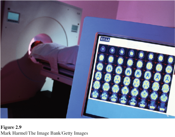
Tools of Discovery—Having Our Head Examined
LOQ 2-
For most of human history, we had no device high-
EEG (electroencephalograph) a device that uses electrodes placed on the scalp to record waves of electrical activity sweeping across the brain’s surface. (The record of those brain waves is an electroencephalogram.)
Now a new generation of mapmakers is at work charting formerly unknown territory, stimulating various brain parts and watching the results. Some use microelectrodes to snoop on the messages of individual neurons. Some attach larger electrodes to the scalp to eavesdrop with an EEG (electroencephalograph) on the chatter of billions of neurons. Others use scans that peer into the thinking, feeling brain and give us a Superman-
PET (positron emission tomography) scan a view of brain activity showing where a radioactive form of glucose goes while the brain performs a given task.
The PET (positron emission tomography) scan tracks a temporarily radioactive form of the sugar glucose. Your brain accounts for only about 2 percent of your body weight. But this control center—
MRI (magnetic resonance imaging) a technique that uses magnetic fields and radio waves to produce computer-
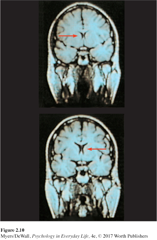
MRI (magnetic resonance imaging) scans capture images of brain structures by briefly disrupting activity in brain molecules. Researchers first position the person’s head in a strong magnetic field, which aligns the spinning atoms of brain molecules. Then, with a brief pulse of radio waves, they disrupt the spinning. When the atoms return to their normal spin, they give off signals that provide a detailed picture of soft tissues, including the brain. MRI scans have revealed, for example, that some people with schizophrenia, a disabling psychological disorder, have enlarged fluid-
fMRI (functional MRI) a technique for revealing bloodflow and, therefore, brain activity by comparing successive MRI scans. fMRI scans show brain function.
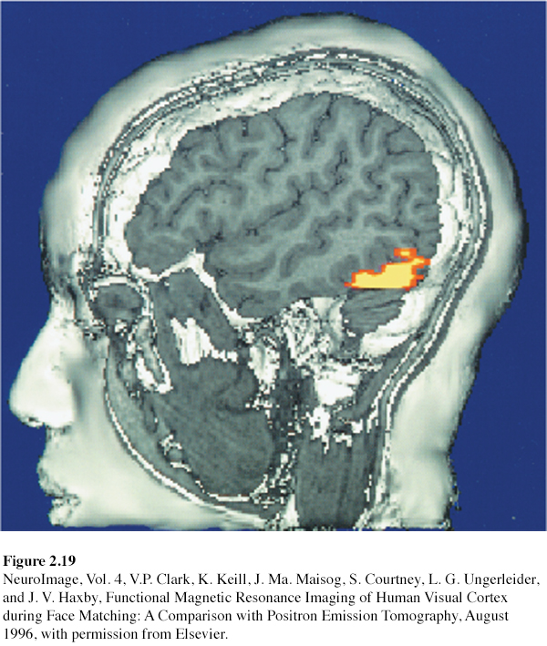
A special application of MRI, fMRI (functional MRI), also reveals the brain’s functions. Where the brain is especially active, blood goes. By comparing MRI scans taken less than a second apart, researchers can watch parts of the brain activate as a person thinks or acts in certain ways. As the person looks at a photo, for example, the fMRI shows blood rushing to the back of the brain, which processes visual information (see FIGURE 2.19). This technology enables a very crude sort of mind reading. Neuroscientists scanned 129 people’s brains as they did eight different mental tasks (such as reading, gambling, and rhyming). Later, viewing another person’s brain images, they were able, with 80 percent accuracy, to identify which of these mental tasks the person was doing (Poldrack et al., 2009).
What the telescope did for astronomy, these brain-
Retrieve + Remember
Question 2.11
•Match the scanning technique with the correct description.
Technique
fMRI scan
PET scan
MRI scan
Description
tracks radioactive glucose to reveal brain activity.
tracks successive images of brain tissue to show brain function.
uses magnetic fields and radio waves to show brain anatomy.
ANSWERS: 1. b, 2. a, 3. c
Older Brain Structures
Brain structures determine our abilities. In sharks and other primitive vertebrates (animals with backbones), a not-

The brain’s increasing complexity arises from new systems built on top of the old, much as new layers cover old ones in Earth’s landscape. Digging down, one discovers the fossil remnants of the past—
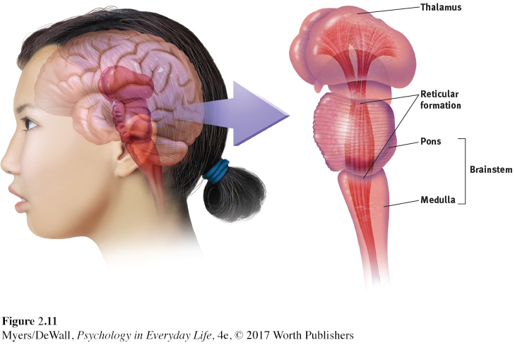
The Brainstem
LOQ 2-
brainstem the oldest part and central core of the brain, beginning where the spinal cord swells as it enters the skull; responsible for automatic survival functions.
medulla [muh-
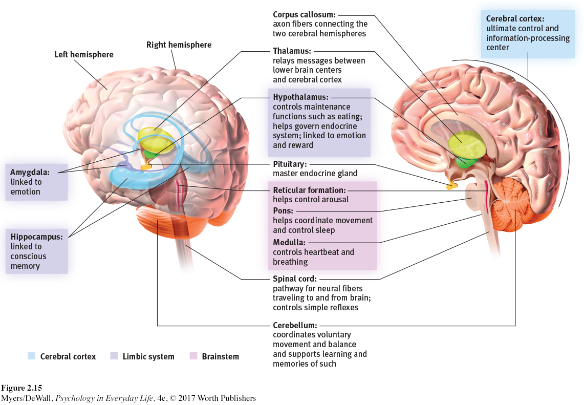
The brainstem is the brain’s oldest and innermost region. Its base is the medulla, the slight swelling in the spinal cord just after it enters the skull (FIGURE 2.11). Here lie the controls for your heartbeat and breathing. Just above the medulla sits the pons, which helps coordinate movements and control sleep. As some severely brain-
The brainstem is a crossover point. Here, you’ll find a peculiar sort of cross-
Retrieve + Remember
Question 2.12
•The _______ is a crossover point where nerves from the left side of the brain are mostly linked to the right side of the body, and vice versa.
ANSWER: brainstem
The Thalamus
thalamus [THAL-
Sitting at the top of the brainstem is the thalamus, which acts as the brain’s sensory control center. This joined pair of egg-
The Reticular Formation
reticular formation nerve network running through the brainstem and into the thalamus; plays an important role in controlling arousal.
Inside the brainstem, between your ears, lies your reticular (“netlike”) formation. This neuron network extends upward from your spinal cord, through your brainstem, and into your thalamus (see FIGURE 2.11). As sensory messages travel from your spinal cord to your thalamus, this long structure acts as a filter, relaying important information to other brain areas.
The reticular formation also controls arousal, as researchers discovered in 1949. Electrically stimulating the reticular formation of a sleeping cat almost instantly produced an awake, alert animal (Moruzzi & Magoun, 1949). When a cat’s reticular formation was cut off from higher brain regions, without damaging nearby sensory pathways, the effect was equally dramatic. The cat lapsed into a coma and never woke up.
The Cerebellum
cerebellum [sehr-
At the rear of the brainstem is the cerebellum, meaning “little brain,” which is what its two wrinkled halves resemble (FIGURE 2.13). This baseball-
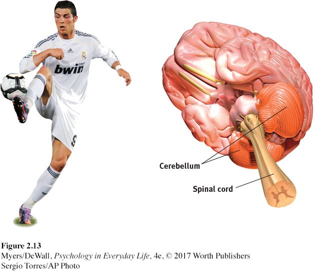
If you answered those questions easily, thank your cerebellum. It helps you judge time, discriminate textures and sounds, and control your emotions (Bower & Parsons, 2003). Your cerebellum also coordinates voluntary movement. When a soccer player masterfully controls the ball, give the cerebellum some credit. If you injured your cerebellum or drugged it with alcohol, you would have trouble walking, keeping your balance, or shaking hands. The cerebellum also helps process and store memories for things you cannot consciously recall, such as how to ride a bicycle. (Stay tuned for more about this in Chapter 7.)
* * *
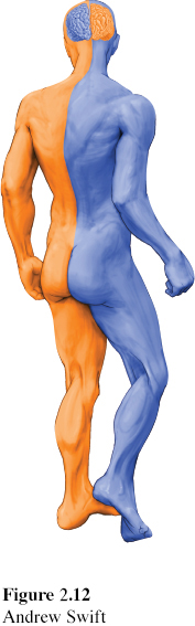
Note: These older brain functions all occur without any conscious effort. Once again, we see one of our Big Ideas at work: Our two-
Retrieve + Remember
Question 2.13
•In what brain region would damage be most likely to (1) disrupt your ability to skip rope? (2) disrupt your ability to hear? (3) perhaps leave you in a coma? (4) cut off the very breath and heartbeat of life?
ANSWERS: 1. cerebellum, 2. thalamus, 3. reticular formation, 4. medulla
The Limbic System
LOQ 2-
limbic system neural system (including the amygdala, hypothalamus, and hippocampus) located below the cerebral hemispheres; associated with emotions and drives.
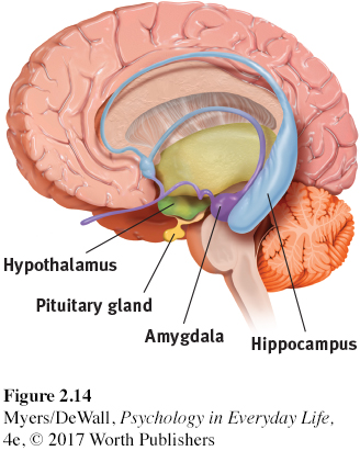
We’ve traveled through the brain’s oldest parts, but we’ve not yet reached its newest and highest regions, the cerebral hemispheres (the two halves of the brain). Before we do, we must pass the limbic system, which lies between the oldest and newest brain areas (limbus means “border”). The limbic system contains the amygdala, the hypothalamus, and the hippocampus (FIGURE 2.14).
amygdala [uh-
THE AMYGDALA The amygdala—two lima-
What, then, might happen if we electrically stimulated the amygdala of a normally mellow domestic animal, such as a cat? Do so in one spot and the cat prepares to attack, hissing with its back arched, its pupils wide, its hair on end. Move the electrode only slightly within the amygdala, cage the cat with a small mouse, and now it cowers in terror.
Many experiments have confirmed the amygdala’s role in processing emotions and perceiving rage and fear. Monkeys and humans with amygdala damage become less fearful of strangers (Harrison et al., 2015). After her amygdala was destroyed by a rare genetic disease, one woman no longer experienced fear. Facing a snake, speaking in public, even being threatened with a gun—
But a critical thinker should be careful here. The brain is not neatly organized into structures that reflect specific behaviors and feelings. When we feel afraid or act aggressively, many areas of our brain become active, not just the amygdala. If you destroy a car’s battery, you won’t be able to start the engine. Yet the battery is merely one link in the whole working system.
Retrieve + Remember
Question 2.14
•Electrical stimulation of a cat’s amygdala provokes angry reactions, suggesting the amygdala’s role in aggression. Which ANS division is activated by such stimulation?
ANSWER: the sympathetic nervous system
hypothalamus [hi-
THE HYPOTHALAMUS Just below (hypo) your thalamus is your hypothalamus, an important link in the command chain that helps your body maintain a steady internal state. Some neural clusters in the hypothalamus influence hunger. Others regulate thirst, body temperature, and sexual behavior.
To monitor your body state, the hypothalamus tunes in to your blood chemistry and any incoming orders from other brain parts. For example, picking up signals from your brain’s information-
A remarkable discovery about the hypothalamus illustrates how progress in science often occurs—
Animal researchers have discovered similar reward centers in or near the hypothalamus in goldfish, dolphins, monkeys, and other species. One general reward system triggers the release of the neurotransmitter dopamine. Specific centers help us enjoy the pleasures of eating, drinking, and sex. Animals, it seems, come equipped with built-
Do we humans also have limbic centers for pleasure? Some evidence indicates we do. To calm violent patients, one neurosurgeon implanted electrodes in such limbic system areas. Although those patients reported mild pleasure, they—
hippocampus a neural center located in the limbic system; helps process for storage explicit (conscious) memories of facts and events.
THE HIPPOCAMPUS The hippocampus—a seahorse-
Later in this chapter, we’ll see how the hippocampus helps store the day’s experiences while we sleep. In Chapter 7, we’ll explore how the hippocampus interacts with our frontal lobes to create our conscious memory.
* * *
FIGURE 2.15 locates the brain areas we’ve discussed—
Retrieve + Remember
Question 2.15
•What are the three key structures of the limbic system, and what functions do they serve?
ANSWER: (1) The amygdala is involved in aggression and fear responses. (2) The hypothalamus is involved in bodily maintenance, pleasurable rewards, and control of the hormonal systems. (3) The hippocampus processes memory of facts and events.
The Cerebral Cortex
cerebral [seh-
Older brain networks sustain basic life functions and enable memory, emotions, and basic drives. High above these older structures are the cerebrum—
Structure of the Cortex
LOQ 2-
If you opened a human skull, exposing the brain, you would see a wrinkled organ, shaped somewhat like an oversized walnut. Without these wrinkles, a flattened cerebral cortex would require triple the area—
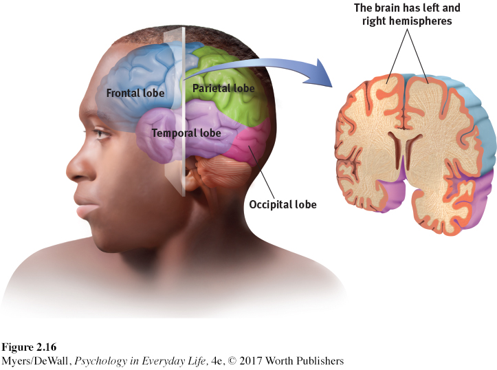
frontal lobes the portion of the cerebral cortex lying just behind the forehead; involved in speaking and muscle movements and in making plans and judgments.
parietal [puh-
occipital [ahk-
temporal lobes the portion of the cerebral cortex lying roughly above the ears; includes areas that receive information from the ears.
Each hemisphere’s cortex is subdivided into four lobes, separated by deep folds (FIGURE 2.16). You can roughly trace the four lobes, starting with both hands on your forehead. The frontal lobes lie directly behind your forehead. As you move your hands over the top of your head, toward the rear, you’re sliding over your parietal lobes. Continuing to move down, toward the back of your head, you’ll slide over your occipital lobes. Now move each hand forward, to the sides of your head, and just above each ear you’ll find your temporal lobes. Each hemisphere has these four lobes. Each lobe carries out many functions. And many functions require the cooperation of several lobes.
Functions of the Cortex
LOQ 2-
More than a century ago, surgeons found damaged areas of the cerebral cortex during autopsies of people who had been partially paralyzed or speechless. This rather crude evidence was interesting, but it did not prove that specific parts of the cortex control complex functions like movement or speech. A laptop with a broken power cord might go dead, but we would be fooling ourselves if we thought we had “localized” the Internet in the cord.
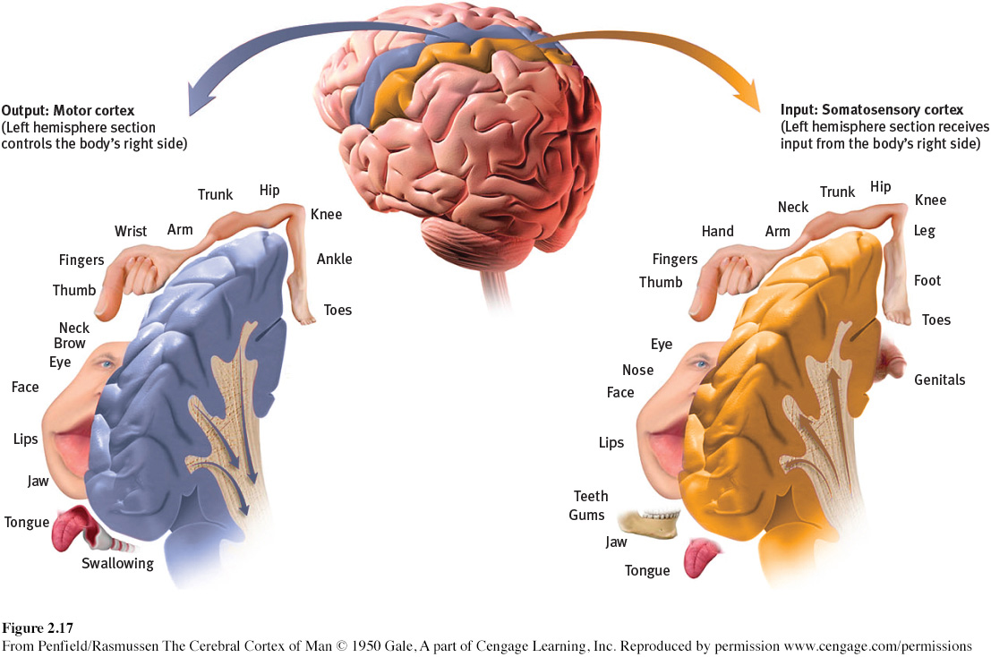
motor cortex the cerebral cortex area at the rear of the frontal lobes; controls voluntary movements.
MOTOR FUNCTIONS Early scientists had better luck showing simple brain-
Lucky for brain surgeons and their patients, the brain has no sensory receptors. Knowing this, in the 1930s, Otfrid Foerster and Wilder Penfield were able to map the motor cortex in hundreds of wide-
As is so often the case in science, new answers have triggered new questions. Might electrodes implanted in the motor cortex identify what neurons control specific activities? If so, could people learn to mentally control these implanted devices, perhaps to direct a robotic limb? Clinical trials are now under way with people who have severe paralysis or have lost a limb (Andersen et al., 2010; Nurmikko et al., 2010). Some who received implants have indeed learned to control robotic limbs (Collinger et al., 2013; Hochberg et al., 2012).
Retrieve + Remember
Question 2.16
•Try moving your right hand in a circular motion, as if polishing a car. Then start your right foot doing the same motion as your hand. Now reverse the right foot’s motion, but not the hand’s. Finally, try moving the left foot opposite to the right hand.
Why is reversing the right foot’s motion so hard?
Why is it easier to move the left foot opposite to the right hand?
ANSWERS: 1. The right limbs’ activities interfere with each other because both are controlled by the same (left) side of your brain. 2. Opposite sides of your brain control your left and right limbs, so the reversed motion causes less interference.
somatosensory cortex the cerebral cortex area at the front of the parietal lobes; registers and processes body touch and movement sensations.
SENSORY FUNCTIONS The motor cortex sends messages out to the body. What parts of the cortex receive incoming messages from our senses of touch and movement? Penfield supplied the answer. An area now called the somatosensory cortex receives this sensory input. It runs parallel to the motor cortex and just behind it, at the front of the parietal lobes (see FIGURE 2.17). Stimulate a point on the top of this band of tissue, and a person may report being touched on the shoulder. Stimulate some point on the side, and the person may feel something on the face.
The more sensitive a body region, the larger the somatosensory area devoted to it. Why do we kiss with our lips rather than rub elbows? Our supersensitive lips project to a larger brain area than do our arms (see FIGURE 2.17). Similarly, rats have a large brain area devoted to their whisker sensations, and owls to their hearing sensations.
Your somatosensory cortex is a very powerful tool for processing information from your skin senses—
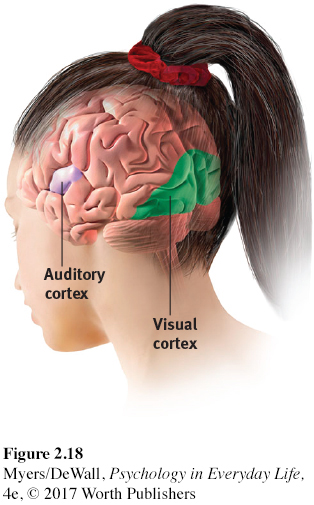
If you have normal vision, you might see flashes of light or dashes of color if stimulated in your occipital lobes. (In a sense, we do have eyes in the back of our head!)
hallucination a false sensory experience, such as hearing something in the absence of an external auditory stimulus.
Any sound you now hear is processed by your auditory cortex in your temporal lobes (just above your ears; see FIGURE 2.18). Most of this auditory information travels a roundabout route from one ear to the auditory receiving area above your opposite ear. If stimulated in your auditory cortex, you alone might hear a sound. People with schizophrenia sometimes have auditory hallucinations (false sensory experiences). MRI scans taken during these hallucinations show active auditory areas in the temporal lobes (Lennox et al., 1999).
Retrieve + Remember
Question 2.17
•Our brain’s _______ cortex registers and processes body touch and movement sensations. The _______ cortex controls our voluntary movements.
ANSWERS: somatosensory; motor
association areas cerebral cortex areas involved primarily in higher mental functions, such as learning, remembering, thinking, and speaking.
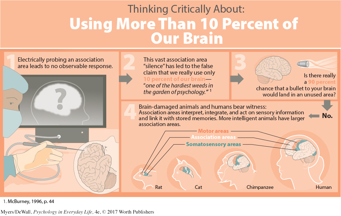
ASSOCIATION AREAS So far, we have pointed out small areas of the cortex that receive messages from our senses, and other small areas that send messages to our muscles. Together, these areas occupy about one-
Association areas are found in all four lobes. In the forward part of the frontal lobes, the prefrontal cortex enables judgment, planning, and processing of new memories. People with damaged frontal lobes may have intact memories, high intelligence test scores, and great cake-
Frontal lobe damage can alter personality, as it did in the famous case study of railroad worker Phineas Gage. One afternoon in 1848, Gage, then 25 years old, was using an iron rod to pack gunpowder into a rock. A spark ignited the gunpowder, shooting the rod up through his left cheek and out the top of his skull, causing massive damage to his frontal lobes (FIGURE 2.20a). To everyone’s amazement, he was immediately able to sit up and speak. After the wound healed, he returned to work. But friendly, soft-
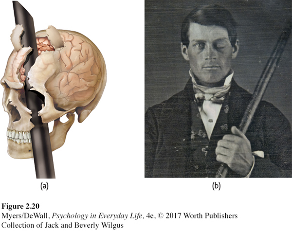
Without the frontal lobe brakes on his impulses, Gage became less inhibited. When his frontal lobes ruptured, his moral compass seemed to disconnect from his behavior. Studies of others with damaged frontal lobes reveal similar losses—
LOQ 2-
Damage to association areas in other lobes would result in different losses. If a stroke or head injury destroyed part of your parietal lobes, you might lose mathematical and spatial reasoning (Ibos & Freedman, 2014). If the damaged area was on the underside of the right temporal lobe, which lets you recognize faces, you would still be able to describe facial features and to recognize someone’s sex and approximate age. Yet you would be strangely unable to identify the person as, say, Taylor Swift or even your grandmother.
Nevertheless, brain scans show that complex mental functions don’t reside in any one spot in your brain. Performing simple tasks may activate tiny patches of your brain, far less than 10 percent. During a complex task, a scan would show many islands of brain activity working together—
Retrieve + Remember
Question 2.18
•Why are association areas important?
ANSWER: Association areas are involved in higher mental functions—
The Power of Plasticity: Responding to Damage
LOQ 2-
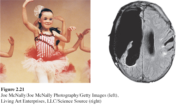
Earlier, we learned about the brain’s plasticity—
Brain-
But there is good news: The brain often attempts self-
neurogenesis the formation of new neurons.
Although self-
Might new drugs spur the production of new nerve cells? Right now, companies are working on such possibilities, so stay tuned. In the meantime, we can all benefit from natural aids to neurogenesis, such as exercise, sex, and sleep (Iso et al., 2007; Leuner et al., 2010; Pereira et al., 2007; Stranahan et al., 2006).
Our Divided Brain
LOQ 2-
Our brain’s look-
Does this mean that the right hemisphere is just along for the ride—
Splitting the Brain: One Skull, Two Minds
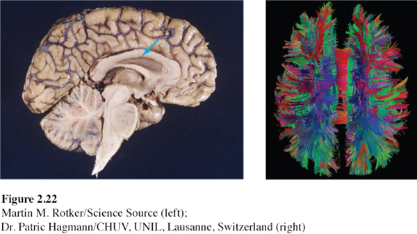
corpus callosum [KOR-
In 1961, two neurosurgeons believed that the uncontrollable seizures of some patients with severe epilepsy was caused by abnormal brain activity bouncing back and forth between the two cerebral hemispheres. If so, they wondered, could they end this biological tennis game by cutting through the corpus callosum, the wide band of axon fibers connecting the two hemispheres and carrying messages between them (FIGURE 2.22)? The neurosurgeons knew that psychologists Roger Sperry, Ronald Myers, and Michael Gazzaniga had divided cats’ and monkeys’ brains in this manner, with no serious ill effects.
split brain a condition in which the brain’s two hemispheres have been isolated by surgery that cut the fibers (mainly those of the corpus callosum) connecting them.
So the surgeons operated. The result? The seizures all but disappeared. The patients with these split brains were surprisingly normal, their personality and intellect hardly affected. Waking from surgery, one even joked that he had a “splitting headache” (Gazzaniga, 1967). By sharing their experiences, these patients have greatly expanded our understanding of interactions between the intact brain’s two hemispheres.
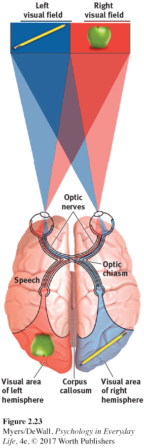
To appreciate these studies, we need to focus for a minute on the peculiar nature of our visual wiring, illustrated in FIGURE 2.23. Note that each eye receives sensory information from the entire visual field. But information from the left half of your field of vision goes to your right hemisphere, and information from the right half of your visual field goes to your left hemisphere. In an intact brain, data received by either hemisphere are quickly transmitted to the other side, across the corpus callosum.
In a person with a severed corpus callosum, this information sharing does not take place. Because the surgery had cut the communication lines between the patient’s two hemispheres, Sperry and Gazzaniga were able to quiz each hemisphere separately. They could send information to the patient’s left hemisphere by having the person stare at a dot and by then flashing a stimulus (a word or photo) to the right of the dot. To send a message to the right hemisphere, they would flash the item to the left of the dot. (If they tried to do this with you, the hemisphere receiving the information would instantly pass the news to the other side of your intact brain.)
In an early experiment, Gazzaniga (1967, 2016) flashed the word HEART across the screen in such a way that HE appeared to the left of the dot, and ART appeared to the right (FIGURE 2.24b). Asked to say what they had seen, the patients reported the letters sent to the left hemisphere, which usually controls speech—
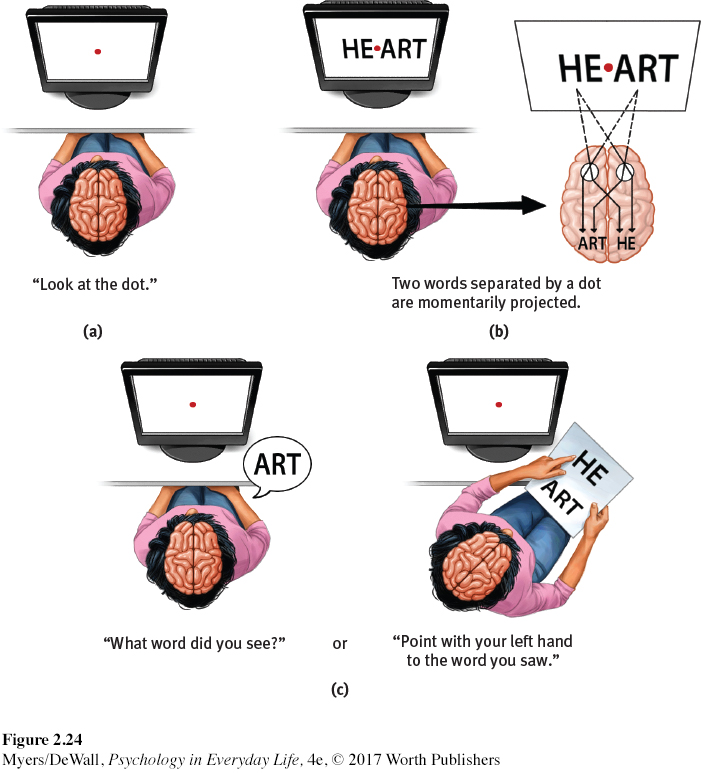
A few people who have had split-
What happens when the “two minds” disagree? If a split-
Retrieve + Remember
Question 2.19
•(1) If we flash a red light to the right hemisphere of a person with a split brain, and flash a green light to the left hemisphere, will each observe its own color? (2) Will the person be aware that the colors differ? (3) What will the person verbally report seeing?
ANSWERS: 1. yes, 2. no, 3. green
Right-Left Differences in Intact Brains
So, what about the 99.99+ percent of us with undivided brains? Does each of our hemispheres also perform distinct functions? The short answer is Yes. If you were performing a perceptual task, a brain scan would show increased activity (brain waves, bloodflow, and glucose consumption) in your right hemisphere. If you were speaking or doing math calculations, the scan would show increased activity in your left hemisphere.
A dramatic demonstration of lateralization happens before some types of brain surgery. To locate the patient’s language centers, the surgeon injects a sedative into the neck artery feeding blood to the left hemisphere, which usually controls speech. Before the injection, the patient is lying down, arms in the air, chatting with the doctor. Can you predict what probably happens when the drug puts the left hemisphere to sleep? Within seconds, the patient’s right arm falls limp. If the left hemisphere is controlling language, the patient will be speechless until the drug wears off.
To the brain, language is language, whether spoken or signed. Just as hearing people usually use the left hemisphere to process spoken language, deaf people usually use the left hemisphere to process sign language (Corina et al., 1992; Hickok et al., 2001). Thus, a left-
Let’s not forget that our left and right brain hemispheres work together. The left hemisphere is good at making quick, exact interpretations of language. But the right hemisphere excels in making inferences (reasoned conclusions) (Beeman & Chiarello, 1998; Bowden & Beeman, 1998; Mason & Just, 2004). It also helps fine-
Simply looking at the two hemispheres, so alike to the naked eye, who would suppose they each contribute uniquely to the harmony of the whole? Yet a variety of observations—