Secondary Structures of DNA
The secondary structure of DNA refers to its three-
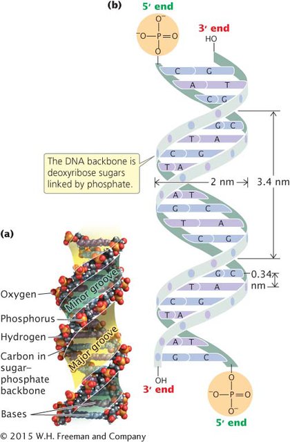
THE DOUBLE HELIX A fundamental characteristic of DNA’s secondary structure is that it consists of two polynucleotide strands wound around each other: it’s a double helix. The sugar–phosphate linkages are on the outside of the helix, and the bases are stacked in the interior of the molecule (see Figure 8.11). The two polynucleotide strands run in opposite directions: they are antiparallel, which means that the 5′ end of one strand is opposite the 3′ end of the other strand.
The strands are held together by two types of molecular forces. Hydrogen bonds link the bases on opposite strands (see Figure 8.11). These bonds are relatively weak compared with the covalent phosphodiester bonds that connect the sugar and phosphate groups of adjoining nucleotides on the same strand. As we will see, several important functions of DNA require the separation of its two nucleotide strands, and this separation can be readily accomplished because of the relative ease of breaking and reestablishing the hydrogen bonds.
The nature of the hydrogen bond imposes a limitation on the types of bases that can pair. Adenine normally pairs only with thymine through two hydrogen bonds, and cytosine normally pairs only with guanine through three hydrogen bonds (see Figure 8.11). Because three hydrogen bonds form between C and G and only two hydrogen bonds form between A and T, C–G pairing is stronger than A–T pairing. The specificity of the base pairing means that wherever there is an A on one strand, there must be a T in the corresponding position on the other strand, and wherever there is a G on one strand, a C must be on the other. The two polynucleotide strands of a DNA molecule are therefore not identical, but rather complementary DNA strands. The complementary nature of the two nucleotide strands provides for efficient and accurate DNA replication (as we will see in Chapter 9).
The second force that holds the two DNA strands together is the interaction between the stacked base pairs in the interior of the molecule. Stacking means that adjacent bases are aligned so that their rings are parallel and stack on top of one another. The stacking interactions stabilize the DNA molecule but do not require that any particular base follow another. Thus, the base sequence of the DNA molecule is free to vary, allowing DNA to carry genetic information.  TRY PROBLEMS 25 AND 27
CONCEPTS
DNA consists of two polynucleotide strands. The sugar phosphate groups of each polynucleotide strand are on the outside of the molecule, and the bases are in the interior. Hydrogen bonding joins the bases of the two strands: guanine pairs with cytosine, and adenine pairs with thymine. The two polynucleotide strands of a DNA molecule are complementary and antiparallel.
 CONCEPT CHECK 5
The antiparallel nature of DNA refers to
its charged phosphate groups.
the pairing of bases on one strand with bases on the other strand.
the formation of hydrogen bonds between bases from opposite strands.
the opposite direction of the two strands of nucleotides.
d
DIFFERENT SECONDARY STRUCTURES As we have seen, DNA normally consists of two polynucleotide strands that are antiparallel and complementary (exceptions are the single-
The three-
B-
Another secondary structure that DNA can assume is the A-
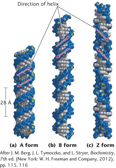
A radically different secondary structure, called Z-
CONCEPTS
DNA can assume different secondary structures, depending on the conditions in which it is placed and on its base sequence. B-
CONCEPT CHECK 6
How does Z-
Z-