CONCEPT14.3 Spatial Differences in Gene Expression Lead to Morphogenesis
Pattern formation is the developmental process that results in the spatial organization of a tissue or organism. An example is the development of the vulva in C. elegans (see Figure 14.8). Pattern formation is inextricably linked to morphogenesis, the development of body form. Underlying both of these processes are spatial differences in gene expression, which determine whether, for example, a particular piece of tissue will become a leg or a wing or a flower petal. These instances where different genes are expressed in different places in the developing organism, in turn, depend on two cellular processes:
- The cells in the tissue must “know” where they are in relation to rest of the body.
- The cells must activate the pattern of gene expression that is appropriate for their location.
In the sections that follow, we will explore the mechanisms used by various organisms to direct pattern formation and morphogenesis.
Morphogen gradients provide positional information during development
During development, the key cellular question “What am I (or what will I be)?” is often answered in part by “Where am I?” Think of the cells in the developing nematode, which develop into different parts of the vulva depending on their positions relative to the anchor cell. The same is true for the cells between the digits of a developing hand and in different whorls of a developing flower. This spatial “sense” is called positional information.
Positional information often comes in the form of an inducer called a morphogen, which diffuses from one group of cells to surrounding cells, setting up a concentration gradient. There are two requirements for a signal to be considered a morphogen:
- It must specifically affect target cells.
- Different concentrations of the signal must cause different effects.
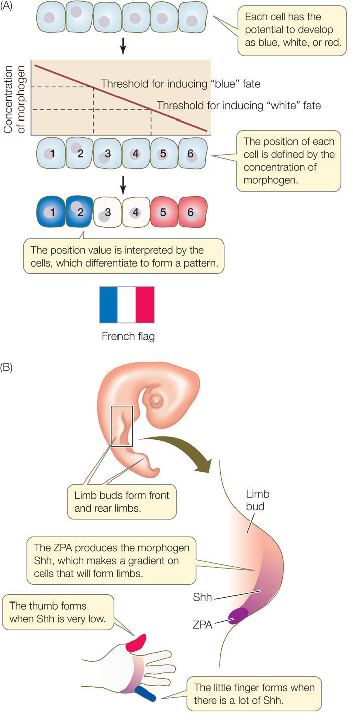
Developmental biologist Lewis Wolpert uses the “French flag model” to explain the action of morphogens (FIGURE 14.11A). This model can be applied to the differentiation of the vulva in C. elegans (see Figure 14.8), which relies on a gradient of LIN-3. Another example can be seen in the development of vertebrate limbs.
The vertebrate limb develops from a paddle-shaped limb bud (FIGURE 14.11B). The cells that develop into different digits must receive positional information; if they do not, the limb will not be organized properly (imagine a hand with only thumbs or only little fingers). How do the cells know where they are? A group of cells at the posterior base of the limb bud, just where it joins the body wall, is called the zone of polarizing activity (ZPA). The cells of the ZPA secrete a morphogen called Sonic hedgehog (Shh), which forms a gradient that determines the posterior–anterior (little finger to thumb) axis of the developing limb. The cells getting the highest dose of Shh form the little finger; those getting the lowest dose develop into the thumb. Recall the French flag model when considering the gradient of Shh.
284
Multiple proteins interact to determine developmental programmed cell death
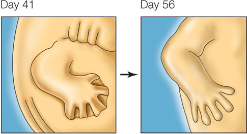
You might expect morphogenesis to involve a lot of cell division, followed by differentiation—and it does. But what you might not expect is the amount of programmed cell death—apoptosis—that occurs during morphogenesis. For example, in an early human embryo, the hands and feet look like tiny paddles: the tissues that will become fingers and toes are linked by connective tissue. Between days 41 and 56 of development, the cells between the digits die, freeing the individual fingers and toes:
Many cells and structures form and then disappear during development, in processes involving apoptosis.
LINK
Concept 7.5 describes some of the cellular events of apoptosis
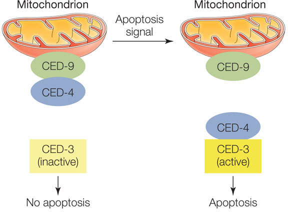
Model organisms have been very useful in studying the genes and proteins involved in apoptosis. For example, the nematode worm C. elegans produces precisely 1,090 somatic cells as it develops from a fertilized egg into an adult, but 131 of those cells die (leaving 959 cells in the adult worm). The sequential activation of two proteins called CED-4 and CED-3 (for cell death) is essential to this programmed cell death. A third protein called CED-9, which is bound to the outside of the mitochondria, inhibits apoptosis in cells that are not programmed to die. In these cells, CED-9 binds CED-4 and prevents it from activating CED-3. If the cell receives a signal for apoptosis, CED-9 releases CED-4, which then activates CED-3:
CED-3 is a caspase (a protease involved in apoptosis) that turns out to be similar to the caspase protein in humans. Several other proteins involved in the nematode apoptosis pathway (including CED-4 and CED-9) also have relatives in humans. So humans and C. elegans, two species separated by more than 600 million years of evolutionary history, have similar genes (encoding similar proteins) that control programmed cell death. The commonality of this pathway indicates its importance: most mutations in the genes that control this pathway are harmful and evolution selects against them. We will return to other examples of links between evolution and development in Concept 14.4.
Our example of apoptosis in the development of fingers and toes shows one of the many ways that the behavior of cells can give rise to body form during development. It also illustrates the two cellular processes underlying pattern formation: only cells in a particular place (between the digits) activate a specific pattern of gene expression (to trigger apoptosis).
Expression of transcription factor genes determines organ placement in plants
Like animals, plants have organs—for example, leaves and roots. Many plants form flowers, and many flowers are composed of four types of organs: sepals, petals, stamens (male reproductive organs), and carpels (female reproductive organs). These floral organs occur in concentric whorls (rings), with groups of each organ type encircling a central axis. The sepals are on the outside and the carpels are on the inside (FIGURE 14.12A).
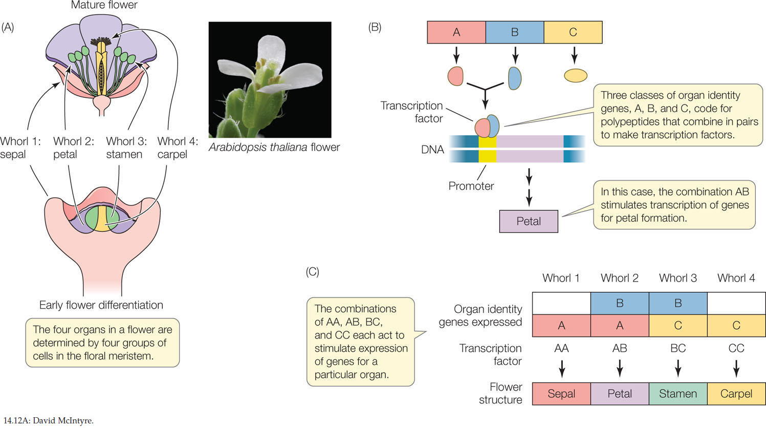
In the model plant Arabidopsis thaliana (thale cress), flowers develop in a radial pattern around the top of the stem as it develops and elongates. At the shoot apex and in other parts of the plant where growth and differentiation occur (such as the root tip), there are groups of undifferentiated, rapidly dividing cells called meristems (see Concept 24.1). Each flower begins as a floral meristem of about 700 undifferentiated cells arranged in a dome, and the four whorls develop from this meristem. How is the identity of a particular whorl determined? Three classes of genes called organ identity genes encode proteins that act in combination to produce specific whorl features (FIGURE 14.12B,C):
- Genes in class A are expressed in whorls 1 and 2 (which form sepals and petals, respectively).
- Genes in class B are expressed in whorls 2 and 3 (which form petals and stamens).
- Genes in class C are expressed in whorls 3 and 4 (which form stamens and carpels).
Gene expression and morphogenesis
Molecular biologists can attach genes to active promoters and insert them into cells. This results in higher than normal expression (overexpression) of the genes. What do you think would happen in each case if the four genes listed were to be overexpressed in the specified tissues? Explain your answers.
- ced-3 in embryonic neuron precursors of C. elegans
- myoD in undifferentiated myoblasts
- Sonic hedgehog in a chick limb bud
- LEAFY in a leaf bud meristem of Arabidopsis
285
These genes encode transcription factors that are active as dimers, that is, proteins with two polypeptide subunits. The composition of the dimer determines which genes the transcription factor activates. For example, a dimer made up of two class A monomers activates transcription of the genes that make sepals; a dimer made up of a class A monomer and a class B monomer results in petals, and so forth.
Two lines of experimental evidence support this model for floral organ determination:
- Loss-of-function mutations: for example, a mutation in a class A gene results in no sepals or petals.
- Gain-of-function mutations: for example, a promoter for a class C gene can be artificially coupled to a class A gene. In this case, the class A gene is expressed in all four whorls, resulting in only sepals and petals. In any organism, the replacement of one organ by another is called homeosis, and this type of mutation is a homeotic mutation.
Transcription of the floral organ identity genes is controlled by other gene products, including the LEAFY protein. Plants with loss-of-function mutations in the LEAFY gene make stems instead of flowers, with increased numbers of modified leaves called bracts. The wild-type LEAFY protein is a transcription factor that stimulates expression of the class A, B, and C genes so that they produce flowers. This finding has practical applications. It usually takes 6–20 years for a citrus tree to produce flowers and fruits. Scientists have made transgenic orange trees expressing the LEAFY gene coupled to a strongly expressed promoter. These trees flower and fruit years earlier than normal trees.
A cascade of transcription factors establishes body segmentation in the fruit fly
A major achievement in studies of developmental biology has been the ever-advancing description of how morphogens act in another model organism, the fruit fly Drosophila melanogaster. As you will see in Concept 14.4, the molecular events that underlie fruit fly development turn out to be similar to events that occur in many other organisms, including ourselves. So they merit examination in some detail.
The insect body is made up of segments that differ from one another. The adult fly has an anterior head (composed of several fused segments), three different thoracic segments, and eight abdominal segments at the posterior end. Each segment gives rise to different body parts: for example, antennae and eyes develop from head segments, wings from the thorax, and so on.
The life cycle of Drosophila from fertilized egg to adult takes about 2 weeks. The egg hatches into a larva, which then forms a pupa, which finally is transformed into the adult fly. By the time a larva appears—about 24 hours after fertilization—there are recognizable segments. The thoracic and abdominal segments all look similar, but the fates of the cells to become different adult segments are already determined.
As with other organisms, fertilization in Drosophila leads to a rapid series of mitoses. However, the first 12 nuclear divisions are not accompanied by cytokinesis. So a multinucleate embryo forms instead of a multicellular embryo. The nuclei are brightly stained in the micrographs below:
286
With no cell membranes to cross, morphogens can diffuse easily within the embryo. As you will see, many of these morphogens affect transcription in the cell nuclei. We focus here on cell fate determination events that occur in the first 24 hours, which were elucidated in Drosophila using genetics:
- First, developmental mutations were identified. For example, a mutant strain might produce larvae with two heads or missing certain segments.
- Second, each mutant was compared with wild-type flies, and the gene responsible for the developmental mistake, and its protein product (if appropriate), was isolated.
- Finally, experiments with the gene (making transgenic flies) and protein (injecting the protein into an egg or embryo) were done to confirm the proposed developmental pathway.
Together, these approaches revealed a sequential pattern (cascade) of gene expression that results in the determination of each segment within 24 hours after fertilization. Several classes of genes are involved:
- Maternal effect genes, which set up the major axes (anterior–posterior and dorsal–ventral) of the egg
- Segmentation genes, which determine the boundaries and polarity of each of the segments
- Hox genes, which determine what organ will be made at a given location
Maternal Effect Genes
Like the eggs and early embryos of many other organisms (see Figure 14.7), Drosophila eggs and larvae are characterized by unevenly distributed cytoplasmic determinants. These molecular determinants, which include both mRNAs and proteins, are the products of specific maternal effect genes. These genes are transcribed in the cells of the mother’s ovary, and the mRNAs are passed to the egg via cytoplasmic bridges. Two maternal effect genes, called bicoid and nanos, help determine the anterior–posterior axis of the egg. (The dorsal–ventral, or back–belly, axis is determined by other maternal effect genes that we will not describe here.)
The mRNAs for bicoid and nanos diffuse from the mother’s cells into what will be the anterior (head) end of the egg. After fertilization, the bicoid mRNA is translated to produce Bicoid protein, a transcription factor that diffuses away from the anterior end, establishing a concentration gradient in the egg cytoplasm:
At this point, the egg is in its multinucleate stage.
Where it is present in sufficient concentration, Bicoid stimulates the transcription of the hunchback gene in the early embryo. Consequently, the nuclei nearest the anterior end are most active in the transcription of the hunchback gene, and the resulting gradient of Hunchback protein (itself a transcription factor) establishes the head, or anterior, region.
Meanwhile, the egg’s cytoskeleton transports the nanos mRNA from the anterior end of the egg to the posterior (tail) end, where it is translated after fertilization. This results in a gradient of the Nanos protein, with the highest concentration at the posterior end. At that end, the Nanos protein inhibits the translation of hunchback mRNA, preventing accumulation of Hunchback protein. Thus the actions of both Bicoid and Nanos establish a Hunchback protein gradient, which determines the anterior and posterior ends of the embryo by influencing the gene expression patterns of the nuclei along the gradient.
The events involving bicoid, nanos, and hunchback begin before fertilization and continue after it, during the multinucleate stage, which lasts a few hours. At this stage the embryo looks like a bunch of indistinguishable nuclei under the light microscope. But the fates of the individual nuclei and the cells they will occupy have already begun to be determined. After the anterior and posterior ends have been established, the next step in pattern formation in fruit flies is the determination of segment number and locations.
Segmentation Genes
The number, boundaries, and polarity of the Drosophila larval segments are determined by proteins encoded by the segmentation genes. These genes are expressed when there are about 6,000 nuclei in the embryo (about 3 hours after fertilization). Three classes of segmentation genes act one after the other to regulate finer and finer details of the segmentation pattern (FIGURE 14.13):
- Gap genes organize broad areas along the anterior–posterior axis. Mutations in gap genes result in gaps in the body plan—the omission of several consecutive larval segments.
- Pair rule genes divide the embryo into units of two segments each. Mutations in pair rule genes result in embryos missing every other segment.
287
- Segment polarity genes determine the boundaries and anterior–posterior organization of the individual segments. Mutations in segment polarity genes can result in segments in which posterior structures are replaced by reversed (mirror-image) anterior structures.
By the end of this part of the cascade, nuclei throughout the embryo “know” which segment they will be part of in the adult fly. The next set of genes in the cascade determines the form and function of each segment.
Hox Genes
Hox genes encode a family of transcription factors that are expressed in different combinations along the length of the embryo, and help determine cell fates within each segment. Expression of certain Hox genes leads to the development of antennae in the head segment, whereas other Hox genes are expressed in the thorax to make wings, and so on. Hox genes are homeotic genes that are shared by most animals, and they are functionally similar to the organ identity genes of plants (see Figure 14.12).
How do we know that the Hox genes determine segment identity? A clue comes from homeotic mutations in Drosophila. A gain-of-function mutation in the Hox gene Antennapedia causes legs to grow on the head in place of antennae (FIGURE 14.14). When another Hox gene, bithorax, is mutated, an extra pair of wings grows in a thoracic segment where wings do not normally occur. So the normal (wild-type) functions of the Hox genes must be to “tell” a segment what organ to form. Hox genes encode transcription factors and have a conserved 180-base-pair sequence called the homeobox (from which the genes get their name). The homeobox encodes a 60-amino acid sequence called the homeodomain. The homeodomain recognizes and binds to a specific DNA sequence in the promoters of its target genes. As you will see in Concept 14.4, this domain is found in transcription factors that regulate development in many other animals with an anterior-posterior axis.
288
LINK
To review transcriptional regulation, see Concept 11.2
CHECKpointCONCEPT14.3
- Outline the steps that determine that a nucleus and cell in the developing Drosophila embryo will be part of an antenna.
- Compare the determination of organ identity in Arabidopsis and Drosophila.
- How does the “French flag model” apply to development in Drosophila?
- In the nematode nervous system, 302 neurons come from 405 precursors. How would you investigate the fate of the 103 “missing” cells? What gene(s) might be involved?
We have seen how positional information leads to changes in the expression of key developmental genes, which in turn control morphogenesis. It turns out that there are remarkable similarities in the genes used to guide development in diverse organisms, and this has led to a new way to look at the evolution of development.