CONCEPT23.2 Some Animal Groups Fall outside the Bilateria
Looking at Figure 23.1, you can see that the protostome and deuterostome animals together comprise a monophyletic group known as the Bilateria. Some major traits that support the monophyly of bilaterians (in addition to genomic analyses) are strong bilateral symmetry, the presence of three distinct cell layers in embryos (triploblasty), and the presence of at least seven Hox genes (see Chapter 14). There are, however, four animal groups—the sponges, ctenophores, placozoans, and cnidarians—that are not bilaterians.
The simplest animals, the sponges, have no distinct tissue types. All other animals groups are usually known as eumetazoans. Two groups treated here as eumetazoans, the ctenophores and placozoans, have weakly differentiated tissue layers (placozoans also lack a nervous system), so some biologists exclude these two groups from Eumetazoa as well. Most eumetazoans have some form of body symmetry, a gut, a nervous system, and tissues organized into distinct organs (although there have been secondary losses of some of these structures in some eumetazoans).
Sponges are loosely organized animals
Although they have some specialized cells, sponges have no distinct embryonic cell layers and no true organs. Early naturalists classified sponges as plants because they were sessile and lacked body symmetry.
476
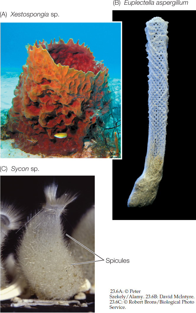
Sponges have hard skeletal elements called spicules, which may be small and simple or large and complex. Three major groups of sponges, which separated soon after the split between sponges and the rest of the animals, are distinguished by their spicules. Members of two groups (demosponges and glass sponges) have skeletons composed of silicaceous spicules made of hydrated silicon dioxide (FIGURE 23.6A,B). These spicules have greater flexibility and toughness than synthetic glass rods of similar length. Members of the third group, the calcareous sponges, take their name from their calcium carbonate spicules (FIGURE 23.6C).
The body plan of all sponges—even large ones, which may reach a meter or more in length—is an aggregation of cells built around a water canal system. Sponges bring water into their bodies by beating the flagella of their specialized feeding cells, the choanocytes (see Figure 23.2B). Water, along with any food particles it contains, enters the sponge by way of small pores and passes into the water canals or a central atrium, where choanocytes capture food particles.
A skeleton of simple or branching spicules, often combined with a complex network of elastic fibers, supports the body of most sponges. Sponges also have an extracellular matrix, composed of collagen, adhesive glycoproteins, and other molecules, that holds the cells together. Most sponges are filter feeders. A few species are carnivores that trap prey on hook-shaped spicules that protrude from the body surface.
477
Most of the 8,500 species of sponges are marine animals. Only about 50 species of sponges live in fresh water. Sponges come in a wide variety of sizes and shapes that are adapted to different movement patterns of water. Sponges living in intertidal or shallow subtidal environments are subject to strong wave action and attach firmly to the substrate. Most sponges that live in slowly flowing water are flattened and are oriented at right angles to the direction of current flow, allowing them to intercept water and the food it contains as it flows past them.
Sponges reproduce both sexually and asexually. In most species, a single individual produces both eggs and sperm, but individuals do not self-fertilize. Water currents carry sperm from one individual to another. Sponges also reproduce asexually by budding and fragmentation.
Ctenophores are radially symmetrical and diploblastic
Ctenophores, also known as comb jellies, were until recently thought to be most closely related to the cnidarians (jellyfishes, corals, and their relatives). But ctenophores lack most of the Hox genes found in all other eumetazoans, and recent studies of their genomes have indicated that ctenophores were among the earliest lineages to split from the remaining animals.
Ctenophores have a radially symmetrical, diploblastic body plan. The two cell layers are separated by an inert, gelatinous extracellular matrix called mesoglea. Ctenophores, unlike sponges, have different openings for food entry and waste elimination: food enters through a mouth, and wastes are eliminated through two anal pores.
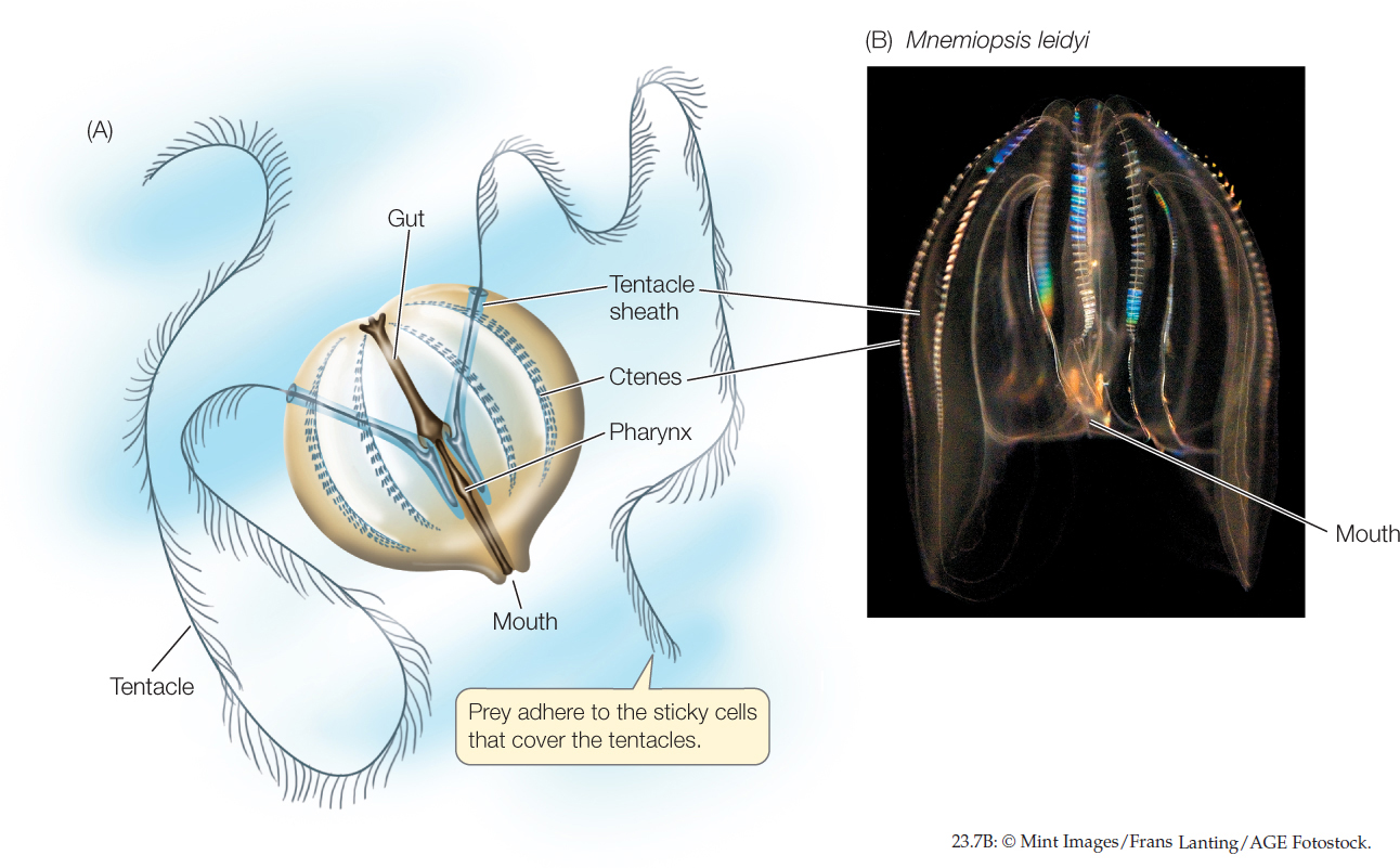
Ctenophores move by beating cilia rather than by muscular contractions. Most of the 250 known species have eight comblike rows of cilia-bearing plates, called ctenes (FIGURE 23.7). The feeding tentacles of ctenophores are covered with cells that discharge adhesive material when they contact prey. After capturing its prey, a ctenophore retracts its tentacles to bring the food to its mouth. In some species, the entire surface of the body is coated with sticky mucus that captures prey. Most ctenophores eat small planktonic organisms, although some eat other ctenophores. Ctenophores are common in open seas and can become abundant in protected bodies of water, where large populations may inhibit the growth of other organisms.
478
Ctenophore life cycles are uncomplicated. Gametes are released into the body cavity and then discharged through the mouth or the anal pores. Fertilization takes place in open seawater. In nearly all species, the fertilized egg develops directly into a miniature ctenophore, which gradually grows into an adult.
Placozoans are abundant but rarely observed
Placozoans are structurally very simple animals with only four distinct cell types (FIGURE 23.8A). Individuals in the mature life stage are usually observed adhering to surfaces (such as the glass of aquariums, where they were first discovered, or to rocks and other hard substrates in nature). Their structural simplicity—they have no mouth, gut, or nervous system—initially led biologists to suspect they might be the sister group of all other animals. Most phylogenetic analyses have not supported this hypothesis, however, and some aspects of the placozoans’ structural simplicity may be secondarily derived. They are generally considered to have a diploblastic body plan, with upper and lower epithelial (surface) layers that sandwich a layer of contractile fiber cells.
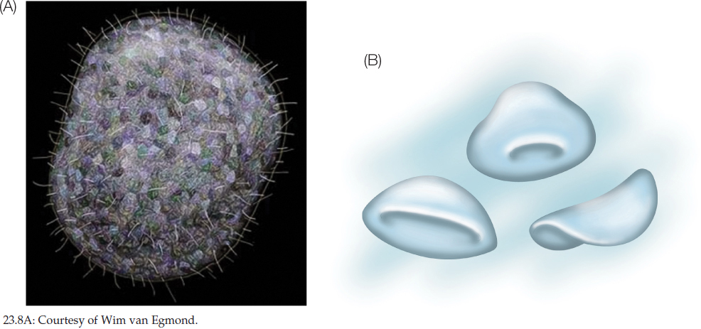
Recent studies have found that placozoans have a pelagic (open-ocean) stage that is capable of swimming (FIGURE 23.8B), but the life history of placozoans is incompletely known. Most studies have focused on the larger adherent stages that can be found in aquariums, where they appear after being inadvertently collected with other marine organisms. The transparent nature and small size of placozoans make them very difficult to observe in nature. Nonetheless, it is known that placozoans can reproduce both asexually as well as sexually, although the details of their sexual reproduction are poorly understood. They are found in warm coastal seas around the world.
Cnidarians are specialized carnivores
The cnidarians (jellyfishes, sea anemones, corals, and hydrozoans) make up the largest and most diverse group of non-bilaterian animals. The mouth of a cnidarian is connected to a blind sac called the gastrovascular cavity. The gastrovascular cavity functions in digestion, circulation, and gas exchange, and it also acts as a hydrostatic skeleton. The single opening serves as both mouth and anus.
The life cycle of many cnidarians has two distinct stages, one sessile and the other motile (FIGURE 23.9), although one or the other of these stages is absent in some groups. In the sessile polyp stage, a cylindrical stalk is attached to the substrate. The motile medusa (plural medusae) is a free-swimming stage shaped like a bell or an umbrella. It typically floats with its mouth and feeding tentacles facing downward.
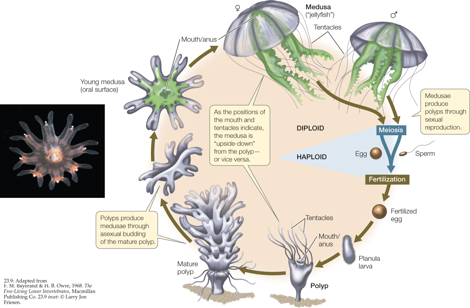
Mature polyps produce medusae by asexual budding. Medusae then reproduce sexually, producing eggs or sperm by meiosis and releasing the gametes into the water. A fertilized egg develops into a free-swimming, ciliated larva called a planula, which eventually settles to the bottom and develops into a polyp.
Cnidarians have epithelial cells with muscle fibers that contract and enable the animals to move, as well as simple nerve nets that integrate their body activities. They are specialized carnivores, using the toxin in harpoonlike structures called nematocysts to capture relatively large and complex prey. Some cnidarians, including many corals and anemones, gain additional nutrition from photosynthetic endosymbionts that live in their tissues.
LINK
The importance of protist endosymbionts in coral colonies is discussed in Concept 20.4
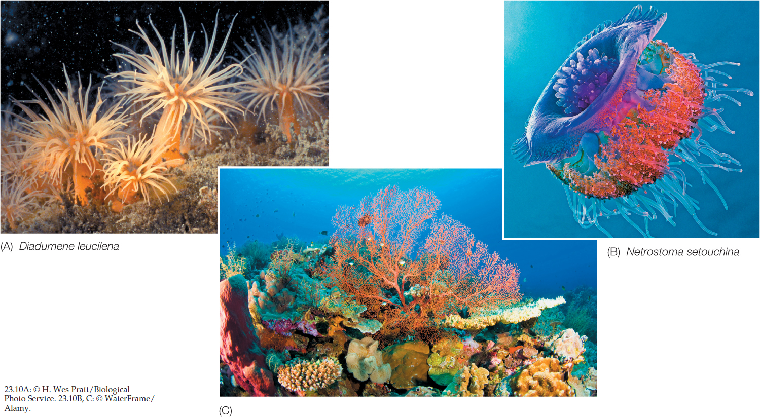
Of the roughly 12,500 living cnidarian species, all but a few live in the oceans. The smallest cnidarians can hardly be seen without a microscope. The largest known jellyfish is 2.5 meters in diameter, and some colonial species can reach lengths in excess of 30 meters. Three clades of cnidarians have many species, some of which are shown in FIGURE 23.10. The anthozoans include sea anemones, sea pens, and corals. The scyphozoans are commonly known as jellyfishes or sea jellies. The less familiar hydrozoans include both freshwater and marine species.
479
480
CHECKpointCONCEPT23.2
- Why are sponges and placozoans considered to be animals, even though they lack the complex body structures found among most other animal groups?
- How does feeding differ between ctenophores and cnidarians?
- Can you think of any advantages to the biphasic life cycle (sessile polyp and motile medusa) of many cnidarians?
In Concept 23.1 we noted that the name protostome means “mouth first,” a developmental synapomorphy that links these animals. The vast majority of animals are protostomes.