CONCEPT32.2 The Breathing Organs and Systemic Tissues Are Usually, but Not Always, in Series
For understanding circulation, we need to recognize two fundamentally different types of tissues or organs in the body of an animal: the systemic tissues and the breathing organs. The systemic tissues are all the tissues and organs of an animal’s body other than the breathing organs (lungs or gills). They take up O2 from the blood and add CO2 to the blood. Conversely, the breathing organs add O2 to the blood and take up CO2 from the blood. With these definitions in mind, we can also define the systemic circuit and the breathing-organ circuit.
The systemic circuit consists of the blood vessels (or blood channels in the case of an open circulatory system) that carry blood to and from the systemic tissues. The breathing-organ circuit consists of the blood vessels that carry blood to and from the breathing organs. This circuit can be called the pulmonary circuit or lung circuit in animals that breathe with lungs, or it can be called the gill circuit in those that breathe with gills.
A final set of important definitions applies to the visible blood vessels, the arteries and veins. These vessels are defined by direction of blood flow, not by the O2 content of the blood they carry. Arteries are vessels that carry blood away from the heart. Veins carry blood toward the heart. Recall from Chapter 31 that perfusion refers to flow of blood through a tissue or organ.
Most fish have the systemic circuit and gill circuit connected in series
The most common circulatory plan in animals is for the systemic and breathing-organ circuits to be connected in series, meaning that blood flows through the two circuits sequentially: first through one circuit, then through the other, then back through the first.
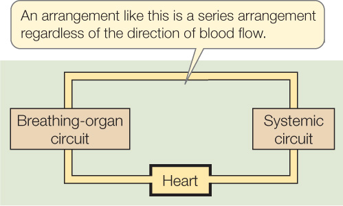
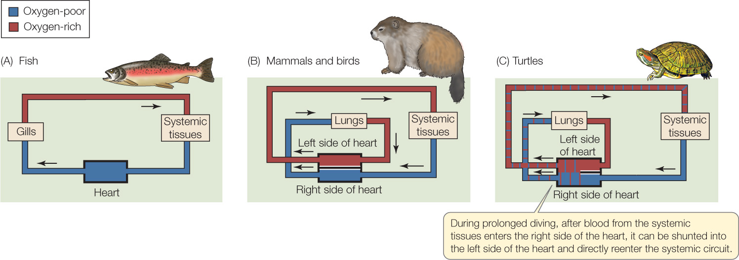
Fish display such a series arrangement (Figure 32.5A). The only exceptions are air-breathing species. In a typical fish, the heart pumps blood into arteries that lead directly to the gills. After the blood perfuses the gills and exits them, it is collected into additional arteries, which branch to supply blood to all the systemic tissues, from the brain in the head to the kidneys and swimming muscles in the abdomen. After perfusing the systemic tissues, the blood enters veins that take it back to the heart.
A series arrangement of this sort is considered to be the most efficient for delivering O2. It is efficient because all the blood pumped to the systemic tissues is O2-rich, having just passed through the gills. Moreover, all the blood pumped to the gills is O2-depleted, having just exited the systemic tissues. Energy is not wasted in sending O2-rich blood to the gills or O2-depleted blood to the systemic tissues.
Natural selection has favored the evolution of a series arrangement in many types of animals. We have already seen, for example, that crustaceans have series circulation in their open circulatory systems (see Figure 32.4). After the blood of a crayfish or crab passes through the gills, the heart pumps it through the systemic circuit, and then the blood returns to the gills. Squid and octopuses, which have closed circulatory systems, also have the gill circuit and systemic circuit connected in series.
666
Mammals and birds have the systemic circuit and lung circuit connected in series
As we will see in more detail in Concept 32.3, mammals and birds have four-chambered hearts, adding complexities to understanding the circulatory plan. However, like fish, they have the systemic circuit and breathing-organ circuit connected in a high-efficiency series arrangement.
In a mammal or bird, blood passes through the heart twice to make a complete trip through the circulatory system. The heart is divided into right and left halves. Moreover, it is completely divided, meaning that sheets of tissue completely separate the right and left halves, so that blood flowing through the right and left sides of the heart cannot mix. As blood travels from the systemic circuit to the lung circuit, it passes through the right half of the heart. As it travels from the lung circuit to the systemic circuit, it passes through the left half of the heart. Figure 32.5B shows the circulatory plan. If you trace the flow of blood from any particular point back to the same point, you will see that blood passes from the systemic circuit to the lung circuit, then back to the systemic circuit, and so forth: a series circulation.
A limitation that fish face is that, after their heart pumps their blood, the blood must pass through both the gill and systemic circuits without receiving additional propulsive energy (an added push) from the heart. Mammals and birds have evolved circulatory plans without this limitation. In a mammal or bird, the heart pumps the blood just before it travels to the lungs, and it pumps it again just before it travels to the systemic circuit.
Amphibians and most non-avian reptiles have circulatory plans that do not anatomically guarantee series flow
What about circulation in the amphibians and non-avian reptiles such as turtles and lizards? Let’s start with a few words about blood flow through the ventricle, the heart’s largest and most muscular chamber. In fish, only O2-depleted blood (from the systemic circuit) enters the ventricle. Thus when the ventricle contracts and ejects blood to flow to the gills, it ejects only O2-depleted blood. In mammals and birds, the ventricle is divided into two anatomically separate chambers (as we will see in Figure 32.6). Only O2-depleted blood enters the right chamber, which then ejects only O2-depleted blood when it contracts. Only O2-rich blood from the lungs enters the left chamber, which therefore pumps exclusively O2-rich blood.
Adult amphibians and most non-avian reptiles have a single ventricle like fish, but blood flow in and out is far more complicated. The lumen of the single ventricle is not a wide-open space because it is occupied by crisscrossing strands of heart muscle and by sheets of connective tissue. Yet the lumen is not fully divided into subparts. O2-rich blood from the lungs enters the ventricle at one point, while simultaneously, O2-depleted blood from the systemic circuit enters it at another point. The ventricle pumps blood to both the lungs and the systemic circuit. In theory, therefore, O2-rich and O2-depleted blood can mix in the ventricle or be pumped to the wrong destination. Decades ago, when biologists knew only about anatomy, the hearts of these animals were often viewed erroneously as being defective intermediate stages in the evolution of vertebrates.
With data from functional studies, we now know that O2-rich and O2-depleted blood are often kept separate as they flow through the ventricle. Investigators have added fluids that can be seen with X-rays to blood of one oxygenation state or the other, for example, and often have observed that little mixing occurs between the O2-rich and O2-depleted blood. In these cases, a series circulation is maintained, with just a small amount of inefficiency as a result of mixing in the ventricle (Figure 32.5C). O2-rich blood from the lungs is pumped by the ventricle principally to the systemic circuit, and O2-depleted blood from the systemic circuit is sent primarily to the lungs.
667
We also now realize from functional studies that sometimes an incompletely divided ventricle can have advantages for certain of these animals. When turtles dive underwater for long periods, for example, their lungs become useless. The turtles, however, have evolved control mechanisms whereby they can stop circulating blood to their lungs. Taking advantage of the fact that they have just a single, undivided ventricle, they direct blood that enters the ventricle from the systemic circuit so that the ventricle pumps it back into the systemic circuit, rather than sending it to the lungs. In this way, the turtles can pump blood round and round in the systemic circuit—permitting the circulatory system to perform all its functions other than gas transport from the lungs—during the time their lungs are useless.
CHECKpointCONCEPT32.2
- What is the systemic circuit?
- Why is a series arrangement of the systemic and breathing-organ circuits efficient for O2 transport?
- In a turtle that has been diving underwater for a long time, does blood pass through the systemic and lung circuits in series? Explain your answer.
By now you have learned a lot about hearts in a general way. But how do hearts work? What controls their regular beating? How do the cells of the heart muscle get the O2 they need? We will address questions such as these in the next concept.