“A STRUCTURE ONLY A TREE WOULD MAKE”
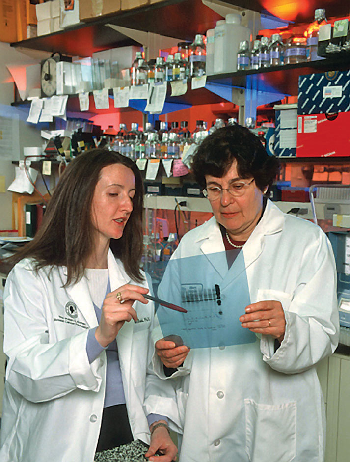
In 1977, Susan Band Horwitz was a young assistant professor working at Albert Einstein College of Medicine when she received a letter from the NCI asking if she would investigate a potential new drug. At the time, Horwitz had never heard of taxol—only one paper had ever been published on it—but as a molecular pharmacologist interested in the chemistry of natural products, she was immediately smitten with taxol’s structure. A strange jumble of rings and tails, it was, she says, “a structure only a tree would make.”
MICROTUBULES Hollow protein fibers that are key components of the cytoskeleton and make up the fibers of the mitotic spindle.
Horwitz and a graduate student, Peter Schiff, gave themselves a month to study the chemical to see if it was worth pursuing further. Within only a few weeks, they knew they had something special. They confirmed that very small quantities of taxol did indeed kill cancer cells growing in a dish. But what really caught their attention was what they saw when they looked at the cancer cells under the microscope. The cells were “loaded,” Horwitz says, with long fibers known as microtubules.
MITOTIC SPINDLE The structure that separates sister chromatids during mitosis.
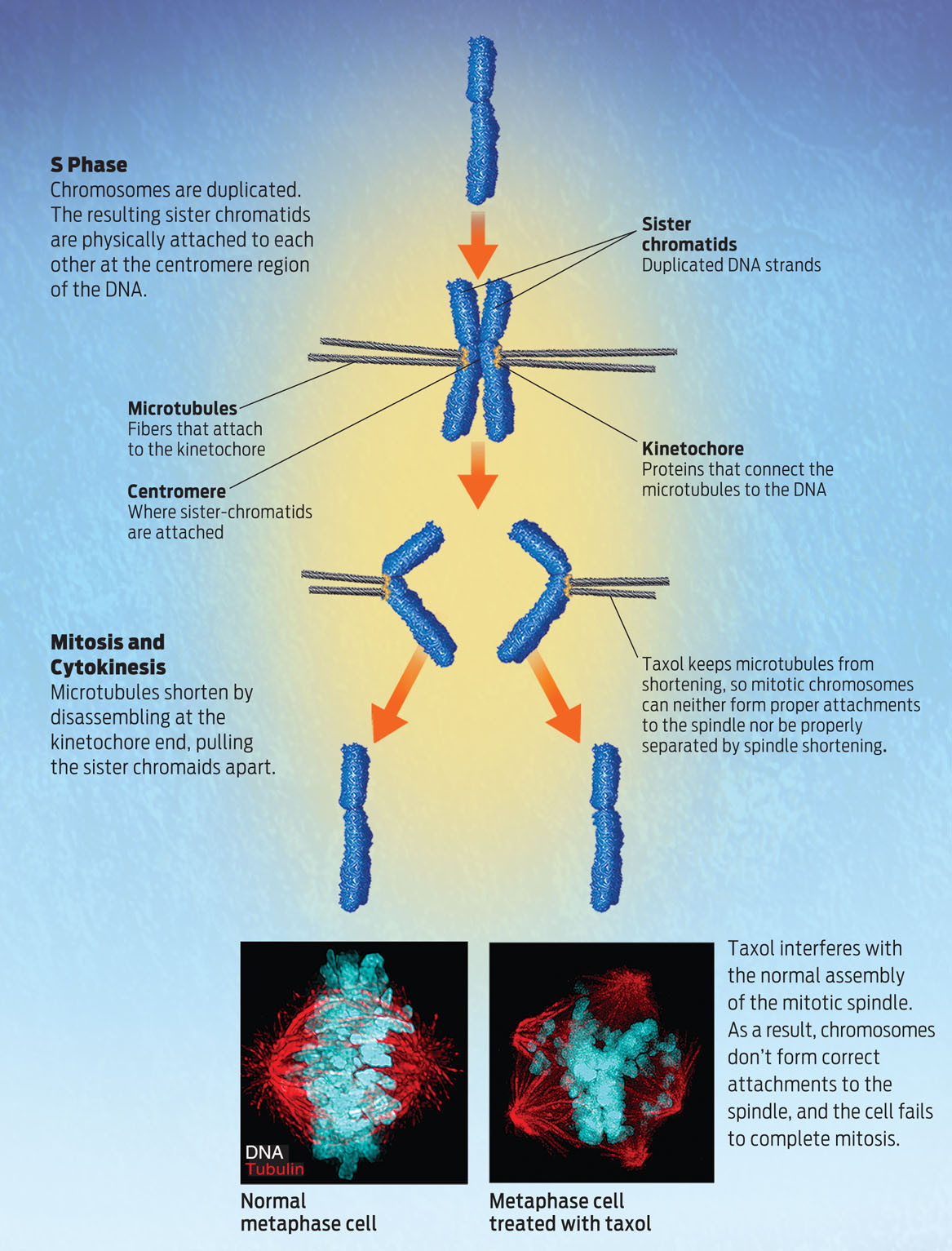
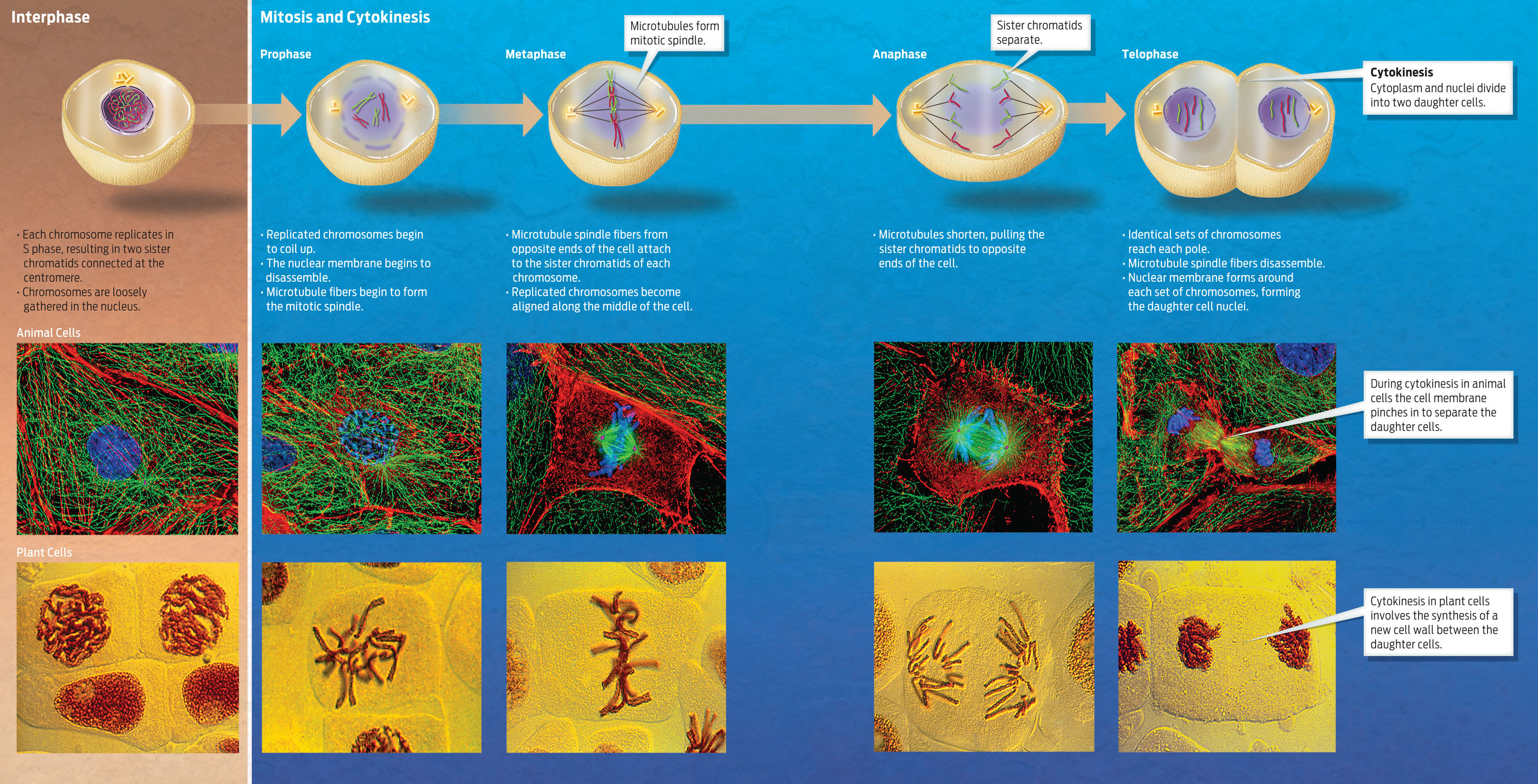
Microtubules are part of the cell’s cytoskeleton (see Chapter 3). Normally, during interphase, microtubules are present as a vast network of fibers running throughout the cytoplasm of the cell. However, as cells enter the early stages of mitosis, the microtubules rearrange to form a structure called the mitotic spindle The microtubules (now known as spindle fibers) begin to attach to sister chromatids of duplicated chromosomes as a cell prepares to enter a phase of mitosis known as metaphase (SEE UP CLOSE: CELL CYCLE AND MITOSIS). The microtubules then tug at the sister chromatids from opposite sides of the cell, pulling the chromatids apart during anaphase. Mitosis is such an important part of the cell cycle that if a cell cannot complete it properly, it self-destructs.
KINETOCHORE Proteins located at the centromere that provide an attachment point for microtubules of the mitotic spindle.
During normal mitosis, microtubules attach to the sister chromatids at the kinetochore, a collection of proteins on the centromere. The microtubules pull the sister chromatids apart by shortening at the end attached to the kinetochore. This shortening involves the disassembly of microtubules at the kinetochore end; subunits are removed from this end of the microtubule, shortening it and dragging the chromatid along with it. The shortening pulls the sister chromatids to opposite ends of the cell. Horwitz and Schiff found that taxol both interferes with the normal organization of microtubules and prevents microtubules from shortening. As a result, the cells are unable to pull sister chromatids apart, and the cells arrest in metaphase (INFOGRAPHIC 9.3).
UP CLOSE: CELL CYCLE AND MITOSIS
No other chemical worked this way, Horwitz says. And that’s what made the discovery so exciting. Existing chemotherapy drugs also targeted the cell cycle, but taxol did it in a new way, which made it a promising new candidate for drug development.