Chapter Introduction

18: Protein Synthesis

617

  • 18.1 The Ribosome

  • 18.2 Activation of Amino Acids for Protein Synthesis

  • 18.3 Initiation of Protein Synthesis

  • 18.4 Elongation and Termination of the Polypeptide Chain

  • 18.5 Translation-Coupled Removal of Defective mRNA

  • 18.6 Protein Folding, Covalent Modification, and Targeting

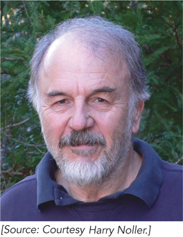
MOMENT OF DISCOVERY

Harry Noller

The defining moment in my career was the realization that ribosomal RNA, not protein, was the functionally important component of the ribosome—and this discovery was somewhat serendipitous. My plan when starting my laboratory, at the University of California, Santa Cruz, was to apply my training in protein biochemistry to investigating the function of ribosomal proteins. We began treating ribosomes with chemicals known to inactivate protein enzymes, hoping to recover nonfunctional ribosomes that could be analyzed to determine which proteins had been affected and thereby discover those responsible for activity. The problem was, the ribosome withstood nearly all the standard chemical treatments we tried!

An unexpected result from one experiment led me to suggest to an undergraduate researcher, Brad Chaires, to try the RNA-specific reagent kethoxal, which reacts with guanine bases to produce adducts that disrupt base pairing. To our great surprise, ribosomes were inactivated by modification of just six G residues out of the more than 4,000 nucleotides in the ribosomal RNA.

When Brad graduated, I followed up with reconstitution experiments showing that it was, in fact, the ribosomal RNA that had been functionally inactivated, and that tRNA could protect ribosomes from kethoxal inactivation. Protection resulted from the tRNA binding to the ribosome and blocking kethoxal’s access to parts of the rRNA, so they couldn’t be chemically modified. These results led us to propose that ribosomal RNA, rather than ribosomal protein, was responsible for the functional activity of the ribosome. Many colleagues considered this “a crackpot idea,” which I found frustrating but, in the antiestablishment spirit of the 1970s, also motivating.

Our findings led us to sequence the ribosomal RNAs (which Carl Woese referred to as “Sacred Scrolls”), and we were excited to find that the sites of chemical inactivation corresponded to the most evolutionarily conserved parts of rRNA. We then embarked on a fruitful collaboration with Woese to determine the secondary structures of the ribosomal RNAs, which ultimately led us in the direction of working out the three-dimensional structure of the ribosome. After almost 40 years, we can at last see that ribosomal RNA is indeed the functional core of the ribosome.

—Harry Noller, on discovering the functional importance of ribosomal RNA

618

The synthesis of proteins is the final step in the flow of genetic information, beginning with DNA replication and continuing with transcription into mRNA. Because of the abundance of proteins—for example, they make up roughly 44% of the dry weight of a human body—and their central catalytic, transport, structural, and regulatory roles in all organisms, substantial cellular resources are devoted to protein synthesis. Like DNA and RNA synthesis, the process of protein synthesis can be considered in terms of initiation, elongation, and termination stages. Furthermore, as in nucleic acid synthesis, substrates must be activated before polymerization, and the completed product—in this case the polypeptide chain—must be chemically modified, folded, and targeted to the correct intracellular or extracellular location to become a functional protein.

The transfer of information from the 4-letter nucleotide sequence of an mRNA to the 20-letter amino acid sequence of a protein, however, is a fundamentally more complex task than nucleotide synthesis. In contrast to the transcription of DNA into RNA, in which a direct correspondence forms by hydrogen bonding between the sequence of the template strand and that of the synthesized RNA strand, there is no obvious chemical correspondence between the three-nucleotide mRNA codons and the amino acids they represent. In the 1950s, Francis Crick suggested that amino acids are attached to “adaptor” RNA molecules that provide direct base-pairing complementarity with each codon in an mRNA sequence. And indeed, Paul Zamecnik and colleagues later showed that amino acids are covalently attached to RNA molecules, subsequently identified as tRNAs (see Chapter 17). The aminoacyl-tRNA molecules associate with the ribosomes, which were shown to be composed of both RNA and protein. Together with mRNAs, tRNAs, and aminoacyl-tRNA synthetases, ribosomes carry out the coupled tasks of recognizing each three-nucleotide codon of the genetic code and incorporating the specified amino acid into a growing polypeptide chain. Experiments in many laboratories showed that the molecular machinery of translation is essentially the same in all cells, although, as we will see, some of the mechanistic details of protein synthesis differ in bacteria and eukaryotes.

In Chapter 17 we introduced the genetic code and the mechanism of decoding by tRNAs. In this chapter, we discuss the structures and activities of ribosomes and aminoacyl-tRNA synthetases. We then explore how these remarkable molecules work together with mRNA and tRNA to carry out fast, accurate protein synthesis.