Highly Specific Enzyme and Antibody Assays Can Detect Individual Proteins
The purification of a protein, or any other molecule, requires a specific assay that can detect the presence of that molecule as it is separated from other molecules (e.g., in column or density-gradient fractions or gel bands or spots). Such an assay capitalizes on some highly distinctive characteristic of a protein: the ability to bind a particular ligand, to catalyze a particular reaction, or to be recognized by a specific antibody. The assay must also be simple and fast to minimize errors and the possibility that the protein of interest will become denatured or degraded while the assay is being performed. The goal of any purification scheme is to isolate sufficient amounts of a given protein for study; thus a useful assay must also be sensitive enough that only a small proportion of the available material is consumed by it. Many common protein assays require just 10−9 to 10−12 g of material.
Chromogenic Enzyme Reactions Many assays are tailored to detect some functional aspect of a protein. For example, assays of enzymatic activity are based on the ability to detect the loss of substrate or the formation of product. Some enzymatic activity assays use chromogenic substrates, which change color in the course of the reaction. (Some substrates are naturally chromogenic; those that are not can be linked to a chromogenic molecule.) Because of the specificity of an enzyme for its substrate, only samples that contain the enzyme will change color in the presence of a chromogenic substrate; the rate of the change provides a measure of the quantity of enzyme present. Enzymes that catalyze chromogenic reactions can also be fused or chemically linked to an antibody and used to “report” the presence or location of an antigen to which the antibody binds (see below).
Antibody Assays As noted earlier, antibodies have the distinctive characteristic of binding tightly and specifically to antigens. As a consequence, preparations of antibodies that recognize a protein antigen of interest can be generated and used to detect the presence of that protein, either in a complex mixture of other proteins (finding a needle in a haystack, as it were) or in a partially or completely purified preparation of a particular protein. The presence of the antigen can be detected by labeling the antibody with an enzyme, a fluorescent molecule, or a radioactive isotope, which can be detected using an enzyme assay, fluorescence microscopy or spectroscopy, or a radiation detector, respectively. For example, luciferase, an enzyme present in fireflies and some bacteria, can be linked to an antibody. In the presence of ATP and its substrate, luciferin, luciferase catalyzes a light-emitting reaction. In either case, after the antibody binds to the protein of interest (the antigen) and unbound antibody is washed away, substrates of the linked enzyme are added and the appearance of color or emitted light is monitored. The intensity is proportional to the amount of enzyme-linked antibody, and thus antigen, in the sample. Alternatively, after a first (or “primary”) antibody that is not otherwise labeled binds to its target protein, a second (“secondary”), labeled antibody that can recognize the first antibody is used to bind to the complex of the first antibody and its target. This combination of primary and secondary antibodies (sometimes called an antibody “sandwich”) permits very high sensitivity in the detection of a target protein because the labeled secondary antibody is often a mixture of antibodies that bind to multiple sites on the first antibody and thus results in a stronger signal than labeling of the primary antibody alone. It is important to remember that an antibody recognizes and binds to only its epitope on a target antigen. If that epitope is altered—for example, by partial unfolding or post-translational modifications—or is blocked when the antigen protein is bound to some other molecule, the ability of the antibody to bind may be reduced or completely lost. Thus the absence of antibody binding does not necessarily mean that the antigen is not present in a sample, only that the epitope portion of that antigen is not present or accessible for antibody binding.
Page 112
To generate antibodies to a protein (discussed in detail in Chapter 23), the intact protein, or a fragment of the protein, is injected into an animal (usually a rabbit, mouse, or goat). Sometimes a short synthetic peptide of 10–15 residues based on the sequence of the protein of interest is used as the antigen to induce antibody formation. Such a synthetic peptide, when coupled to a large protein carrier, can induce an animal to produce antibodies that bind specifically to that part (the epitope) of the full-sized, natural protein. Biosynthetically or chemically attaching the epitope to an unrelated protein is called epitope tagging. As we’ll see throughout this book, antibodies generated using either synthetic peptide epitopes or intact proteins are extremely versatile reagents for isolating, detecting, and characterizing proteins.
Detecting Proteins by Attaching Green Fluorescent Protein An alternative to epitope tagging that is particularly useful in detecting specific proteins within live cells makes use of green fluorescent protein (GFP), a naturally fluorescent protein found in jellyfish (see Figure 4-16). A chimeric protein containing both the protein of interest and GFP, linked together in one polypeptide chain, is expressed in cells by introducing into the cells a gene encoding the combined protein. The amounts and intracellular distribution of the chimeric protein can then be determined readily. This chimeric protein approach is described in Chapter 4.
Detecting Proteins in Gels Proteins embedded within a gel usually are not visible. The two general approaches to detecting proteins in gels are either to label or stain the proteins while they are still within the gel or to electrophoretically transfer the proteins to a membrane made of nitrocellulose or polyvinylidene difluoride and then detect them. Proteins within gels are usually stained with an organic dye or a silver-based stain, both of which are detectable with normal visible light, or with a fluorescent dye, which requires specialized detection equipment. Coomassie blue, the most commonly used organic dye, is typically used to detect about 1000 ng of protein, with a lower limit of detection of about 4–10 ng. Silver staining and fluorescence staining are more sensitive (with a lower limit of ~1 ng). Coomassie and other stains can also be used to visualize proteins after transfer to membranes; however, the most common method of visualizing proteins in membranes is immunoblotting.
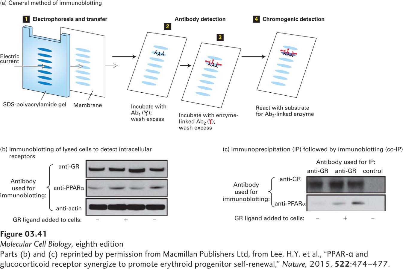
Immunoblotting, also called Western blotting, combines the resolving power of gel electrophoresis with the specificity of antibodies. This multistep procedure is commonly used to separate proteins and then identify a specific protein of interest. As shown in Figure 3-41, two different antibodies are used, one that is specific for the protein of interest (primary antibody) and a secondary antibody that binds to the first and is linked to an enzyme or other molecule that permits detection of the first antibody (and thus the protein of interest to which it binds). The enzyme to which the second antibody is attached can either generate a visible colored product or, by a process called chemiluminescence, produce light that can readily be recorded by film or a sensitive detector. An example of the results of an immunoblotting experiment can be seen in Figure 15-10. If an antibody to the protein of interest is not available, but the gene encoding the protein is available and can be used to express the protein, recombinant DNA methods (see Chapter 5) can incorporate a small peptide epitope into the normal sequence of the protein (epitope tagging) that can be detected by a commercially available antibody to that epitope.
Page 113
[Parts (b) and (c) reprinted by permission from Macmillan Publishers Ltd, from Lee, H.Y. et al., “PPAR-α and glucocorticoid receptor synergize to promote erythroid progenitor self-renewal,” Nature, 2015, 522:474–477.]
EXPERIMENTAL FIGURE 3-41Immunoblotting (IP, or Western blotting) and co-immunoprecipitation (co-IP) can detect specific proteins and their binding partners. (a) Immunoblotting method. Step 1: After a protein mixture has been electrophoresed through an SDS gel, the separated bands (or spots, for two-dimensional gel electrophoresis) are transferred (blotted) from the gel onto a porous membrane from which the protein is not readily removed. Individual proteins (represented by blue ovals) are not visible at this stage. Step 2: The membrane is flooded with a solution of an antibody (Ab1) specific for the protein of interest and allowed to incubate for a while. Ab1 binds to the protein of interest (second from the top), but not to any other proteins attached to the membrane, forming a layer of antibody molecules coincident with the protein (whose position still cannot be seen at this point). Then the membrane is washed to remove unbound Ab1. Step 3: The membrane is incubated with a second antibody (Ab2) that specifically recognizes and binds to the first (Ab1). This second antibody is covalently linked to an enzyme that catalyzes a chromogenic reaction or releases light (e.g., chemiluminescence), a radioactive isotope, or some other substance whose presence can be detected with great sensitivity. Step 4: Finally, the location and amount of bound Ab2 are detected (e.g., by its color for a chromogenic reaction or by detectors or film that measure the light released by chemiluminescence), permitting the electrophoretic mobility (and therefore the mass) of the protein of interest to be determined as well as its quantity (based on band intensity). (b) Immunoblotting was used to detect intracellular receptors and the influence of exposure to a ligand for one of the receptors. In this experiment, cells that are precursors to red blood cells were maintained in vitro in petri dishes and then treated with no ligand (−, leftmost and rightmost lanes) or a ligand that binds to GR, the glucocorticoid receptor (+, center lane). The cells were then lysed in detergent, and immunoblotting (Western blotting) was performed on the total cell lysates using three different antibodies that bind to GR (anti-GR), to a receptor called PPARα (anti-PPARα), or to an abundant intracellular protein, actin, whose presence and abundance was not expected to be sensitive to treatment with the ligand. The equal intensities of the immunoblotting bands detected using the anti-actin antibody (bottom box) provided a “loading control,” which established that essentially equal amounts of cell lysate were applied (loaded) in each lane of the gel. The approximately equal intensities of the bands for both GR and PPARα with or without prior incubation of the cells with the GR ligand showed that the ligand did not substantially alter the amounts of either of these proteins in the cells. Portions of the same cell lysates used for the immunoblotting in part (b) were also used for the immunoprecipitation/immunoblotting shown in part (c). (c) Immunoprecipitation (IP) followed by immunoblotting (together called co-IP) was used to determine if the GR ligand can induce formation of a stable complex that contains both GR and PPARα. Portions of the cell lysates were immunoprecipitated with an antibody to GR (left and center lanes) or a control antibody (right lane) that cannot bind to either GR or PPARα. The immunoprecipitates were separated from the rest of the lysates by centrifugation and then analyzed by immunoblotting with either anti-GR (top box) or anti-PPARα (bottom box). As expected, the top box shows that the GR protein was detected in the immunoprecipitates generated using the α-GR when the same anti-GR antibody was used for the immunoblotting, but not in the immunoprecipitates generated with the control antibody (no band observed). Strikingly, when one examines the immunoprecipitates by immunoblotting with the anti-PPARα antibody (bottom box), a substantial amount of PPARα is seen when the GR ligand is present (center lane), whereas little co-precipitates in the absence of the GR ligand (left lane) or in the control immunoprecipitate (right lane). These results indicate that the GR ligand induces formation of a complex containing both the glucocorticoid receptor and the PPARα proteins. These results do not establish whether or not the GR and PPARα proteins bind directly to each other when the GR ligand is present or if there are additional molecules in the complex that act as intermediates holding the GR and PPARα tightly together when the ligand is present.
[Parts (b) and (c) reprinted by permission from Macmillan Publishers Ltd, from Lee, H.Y. et al., “PPAR-α and glucocorticoid receptor synergize to promote erythroid progenitor self-renewal,” Nature, 2015, 522:474–477.]
ImmunoprecipitationImmunoprecipitation, often abbreviated as IP, exploits the specificity of antibodies to separate a protein of interest from other molecules in a complex mixture—for example, all proteins extracted from a sample of cells or a sample of blood. An antibody to the protein of interest is added to a sample, and the antibody is given time to bind to epitopes on the target protein. An agent that binds to the antibody is then added to cause the antibody and its bound target to precipitate out of solution into particles that can be isolated by centrifugation. A detailed example of this technique is described in Chapter 15 (p. 684). The precipitate is then solubilized under denaturing conditions—for example, in a detergent-containing buffer—to separate the antibody from the protein, and the immunoprecipitated target protein can then be analyzed. If the immunoprecipitated target is tightly bound to one or more other molecules, those bound molecules may be precipitated along with the protein of interest (co-immunoprecipitation, sometimes abbreviated as co-IP). The co-IP method is used frequently to identify and characterize quaternary structures and supramolecular complexes.