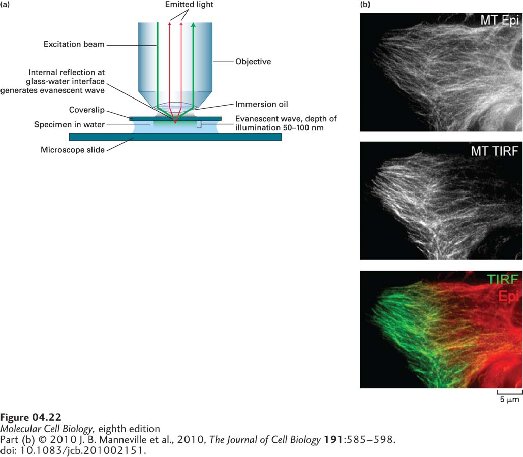
TIRF Microscopy Provides Exceptional Imaging in One Focal Plane
The confocal microscopes we have just described provide amazing and informative images, but they are not perfect. Scientists continue to develop systems that are optimized for special circumstances. Some experimental situations call for fluorescence imaging in a thin focal plane adjacent to a surface, where it would be optimal to minimize out-of-focus background. For example, confocal imaging is not ideal for exploring the details of proteins at adhesion sites between a cell and a coverslip, or for following the kinetics of assembly of microtubules attached to a coverslip. Both of these situations can be imaged at high sensitivity using total internal reflection fluorescence (TIRF) microscopy, in which only the portion of the specimen immediately adjacent to the coverslip is illuminated. In the most common configuration of TIRF microscopy, the excitation light comes through the objective lens (Figure 4-22a). However, the angle at which the light arrives at the coverslip is adjusted so that the light is reflected off the coverslip and returns up through the objective. This generates a narrow band of light, called an evanescent wave, that illuminates only about 50–100 nm of the sample adjacent to the coverslip (2–4 times the thickness of a microtubule), with no illumination of the rest of the sample. Thus if you have a complex mixture of fluorescent structures in a specimen, the TIRF microscope will show you only those that are within 50–100 nm of the coverslip. TIRF has been exceptionally useful in identifying structures on the bottoms of cells grown on a coverslip (Figure 4-22b) and for measuring the kinetics of assembly and disassembly of structures such as microtubules and actin filaments (see Chapters 17 and 18).
EXPERIMENTAL FIGURE 4-22Fluorescent samples in a restricted focal plane can be imaged by total internal reflection fluorescence (TIRF) microscopy. (a) In TIRF microscopy, only about 50–100 nm of the specimen adjacent to the coverslip is illuminated, so that fluorescent molecules in the rest of the sample are not excited. This limited illumination is achieved by directing the illuminating light at an angle at which it is reflected from the glass-water interface of the coverslip rather than passing through it. Whereas most of the light is reflected, it also generates a very small region of illumination called the evanescent wave (depicted in light green). (b) Immunofluorescence microscopy with tubulin antibody was used to visualize microtubules viewed by conventional fluorescence microscopy (top) and by TIRF (middle), and a merged image was created from the two views (bottom). The two images were collected and false-colored red and green so that the merge could highlight those microtubules that are close to the coverslip (green).