CONCEPT39.4 The Adaptive Humoral Immune Response Involves Specific Antibodies
Every day in the human body, billions of B cells survive the test of clonal deletion and are released from the bone marrow into the circulatory system. B cells are the basis for the humoral immune response.
Plasma cells produce antibodies that share a common overall structure
A B cell begins as a “naïve” B cell with a receptor protein on its cell surface that is specific for a particular antigen. The cell is activated by antigen binding to this receptor, and after stimulation by a TH cell, it gives rise to a clone of plasma cells that make antibodies as well as to a smaller number of memory cells (see Figure 39.6). The stimulation occurs after the B cell presents antigen to a TH cell with a receptor that can recognize the antigen. The TH cell then secretes cytokines that stimulate the B cell to divide. This process is described in more detail in Concept 39.5.
All the plasma cells arising from a given B cell produce antibodies that are specific for the antigen that originally bound to the parent B cell. Thus antibody specificity is maintained as B cells proliferate.
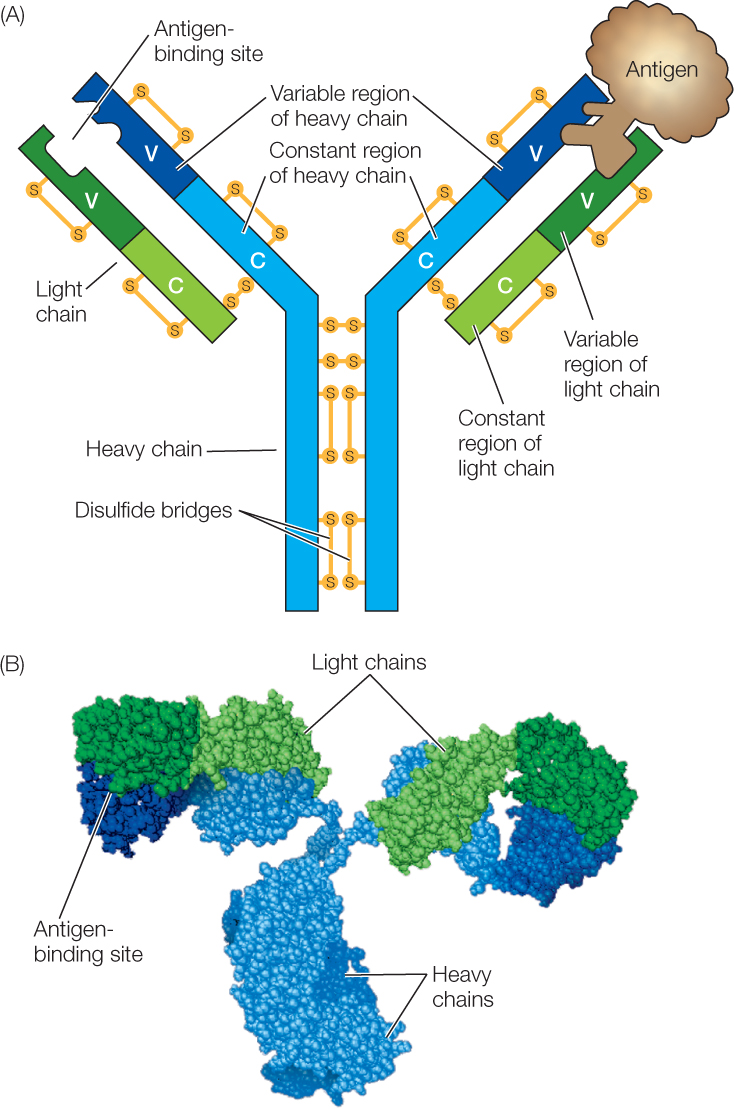
There are several classes of antibodies, also called immunoglobulins, but all contain a tetramer with four polypeptide chains (FIGURE 39.8). In each immunoglobulin molecule, two of the polypeptides are identical light chains, and two are identical heavy chains. Disulfide bridges (see Chapter 3, Concept 3.2) hold the chains together. Each polypeptide chain has a constant region and a variable region:
- The amino acid sequence of the constant region determines the general structure and function (the class) of an immunoglobulin. All immunoglobulins in a particular class have a similar constant region. When an antibody acts as a B cell receptor, the constant region is inserted into the cell membrane, anchoring the antibody to the cell surface.
- The amino acid sequence of the variable region is different for each specific immunoglobulin. The three-dimensional shape of the variable region’s antigen-binding site is responsible for antibody specificity.

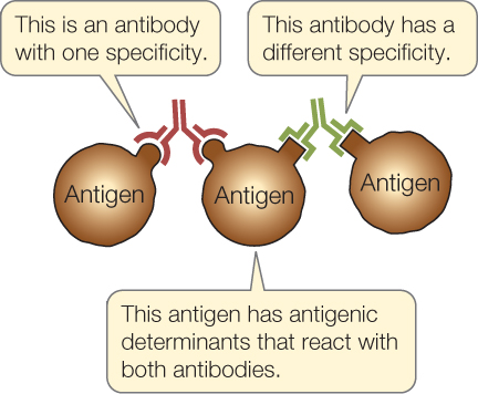
The two antigen-binding sites on each immunoglobulin molecule are identical, making the antibody bivalent (bi, “two”; valent, “binding”). This ability to bind two antigen molecules at once, along with the existence of multiple epitopes (antigenic determinants) on each antigen, permits antibodies to form large complexes with the antigens. For example, one antibody might bind two molecules of an antigen. Another antibody might bind the same antigen at a different epitope or to an antigen that is already bound to the first antibody, along with a third antigen molecule:

820
This binding of multiple antigens and multiple antibodies can result in large complexes that are easy targets for ingestion and breakdown by phagocytes.
There are five classes of immunoglobulins (Ig), and they differ in function and in the type of heavy chain constant region:
- IgG constitutes about 80 percent of circulating antibodies.
- IgD is a cell surface receptor on a B cell.
- IgM is the initial surface and circulating antibody released by a B cell.
- IgA protects mucous membranes exposed to the environment.
- IgE binds to mast cells and is involved with inflammation.
We will focus on IgG antibodies in this chapter.
Antibody diversity results from DNA rearrangements and other mutations
Each mature B cell makes antibodies targeted to only one single antigen. And there are millions of possible antigens to which a human can be exposed. How can the genome encode enough different antibodies to protect the body against all the possible pathogens? Although there are millions of possible amino acid sequences in immunoglobulins, there are not millions of different immunoglobulin genes. It turns out that instead of a single gene encoding each complete immunoglobulin, the genome of the differentiating B cell has multiple different coding regions for each domain of the protein, and diversity is generated by putting together different combinations of these regions. Shuffling of this genetic deck generates the enormous immunological diversity that characterizes each individual mammal.
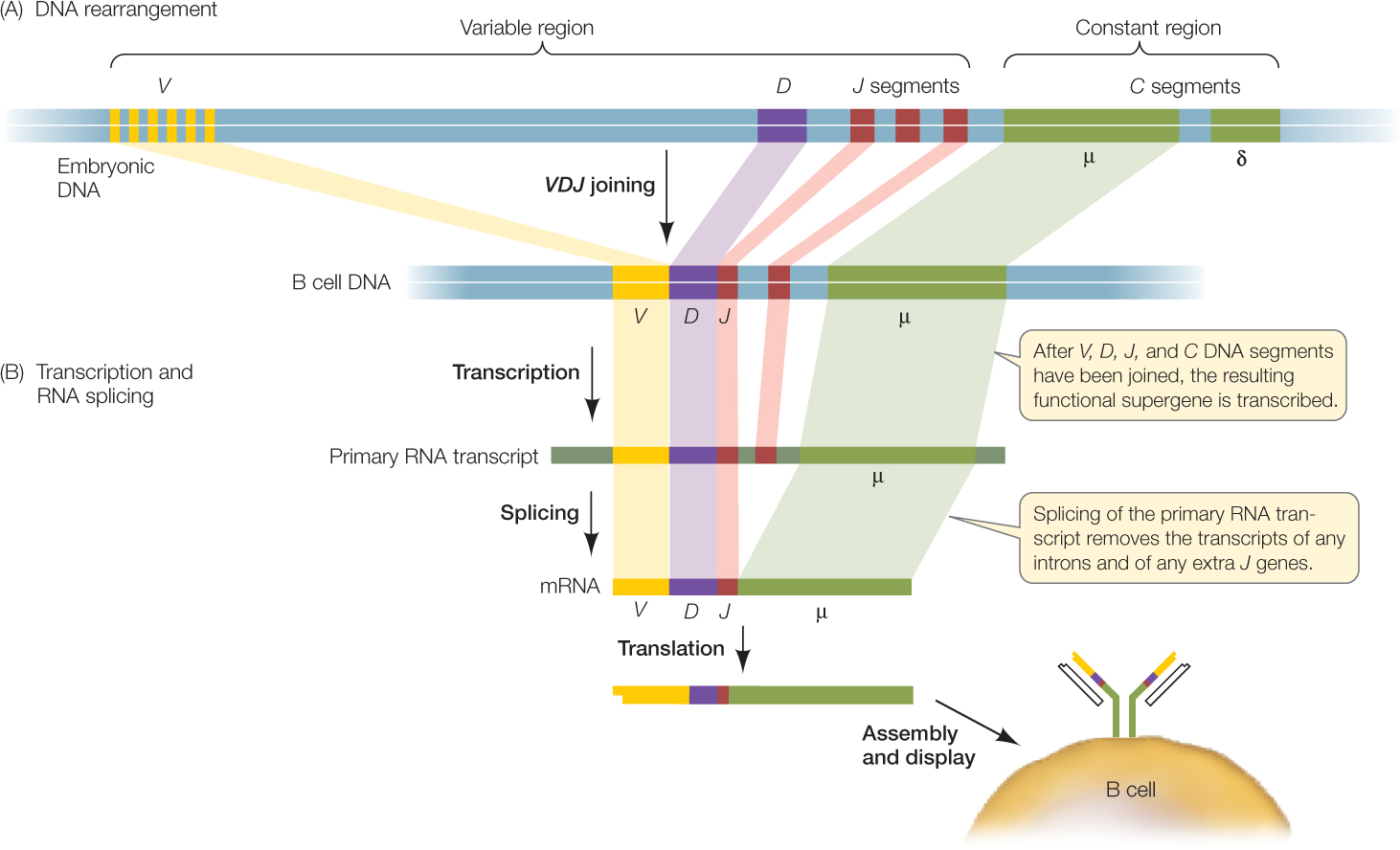
Each gene encoding an immunoglobulin chain is in reality a “supergene” assembled by means of genetic recombination from several clusters of smaller genes scattered along part of a chromosome (FIGURE 39.9). Every cell in the body has hundreds of immunoglobulin genes located in separate clusters that are potentially capable of participating in the synthesis of both the variable and constant regions of immunoglobulin chains. In most body cells and tissues, these genes remain intact and separated from one another. But during B cell development, these genes are cut out, rearranged, and joined together in DNA recombination events. One gene from each cluster is chosen randomly for joining, and the others are deleted. In the case of one multigene set, the J genes, some of the extra sequences are removed by RNA splicing (FIGURE 39.10).


In this manner, a unique immunoglobulin supergene is assembled from randomly selected “parts.” Each B cell precursor assembles two supergenes, one for a specific heavy chain and the other, assembled independently, for a specific light chain. This remarkable example of irreversible cell differentiation generates an enormous diversity of immunoglobulins from the same genome. It is a major exception to the generalization that all somatic cells derived from the fertilized egg have identical DNA.
Figure 39.9 illustrates the gene families that encode the constant and variable regions of the heavy chain in mice. There are multiple genes that encode each of the three parts of the variable region: 100 V, 30 D, and 6 J genes. Each B cell randomly selects one gene from each of these clusters to make the final coding sequence (VDJ) of the heavy-chain variable region. So the number of different heavy chains that can be made through this random recombination process is quite large:
100 V × 30 D × 6 J = 18,000 possible combinations
Now consider that the light chains are similarly constructed, with a similar amount of diversity made possible by random recombination. If we assume that the degree of potential light-chain diversity is the same as that for heavy-chain diversity, the number of possible combinations of light- and heavy-chain variable regions is:
18,000 different light chain × 18,000 different heavy chains = 324 million possibilities!
Other mechanisms generate even more diversity:
- When the DNA sequences that encode the V, D, and J regions are rearranged so that they are next to one another, the recombination event is not precise, and errors occur at the junctions. This imprecise recombination can create frameshift mutations, generating new codons at the junctions, with resulting amino acid changes.
- After the DNA sequences are cut and before they are rejoined, the enzyme terminal transferase often adds some nucleotides to the free ends of the DNA pieces. These additional bases create insertion mutations.
- There is a relatively high spontaneous mutation rate in immunoglobulin genes. Once again, this process creates many new alleles and adds to antibody diversity.
LINK
The effects of DNA mutations on the amino acid sequences of proteins are described in Concept 10.3
When we include these possibilities with the millions of combinations that can be made by random DNA rearrangements, it is not surprising that the immune system can mount a response to almost any natural or artificial substance.
Once the DNA rearrangements are completed, each supergene is transcribed and then translated to produce an immunoglobulin light chain or heavy chain. These chains combine to form an active immunoglobulin protein.
The adaptive humoral immune response involves specific antibodies
Mammals have two kinds of light chains: the kappa (κ) and lambda (λ) chains, and each variable region consists of a V domain and a J domain. Imagine a strain of mouse that has a family of 350 genes encoding the κ V domain and 10 encoding the κ J domain, while the λ variable regions are encoded by 300 V and 5 J genes. The IgG heavy chain is encoded by 350 V, 8 J, and 5 D genes.
- How many different types of antibodies can be made from these genes by recombination?
- What are two other ways to generate antibody diversity from these genes?
Antibodies bind to antigens and activate defense mechanisms
Recall that antibodies have two roles in B cells after they undergo DNA rearrangements and RNA splicing. First, by being expressed on the cell surface, a unique antibody can act as a receptor for an antigen in the recognition phase of the humoral response (see Figures 39.6 and 39.7). Second, in the effector phase of the humoral response, specific antibodies are produced in large amounts by a clone of B cells. These antibodies are secreted from the B cells and enter the bloodstream where they act in either of two ways:
822
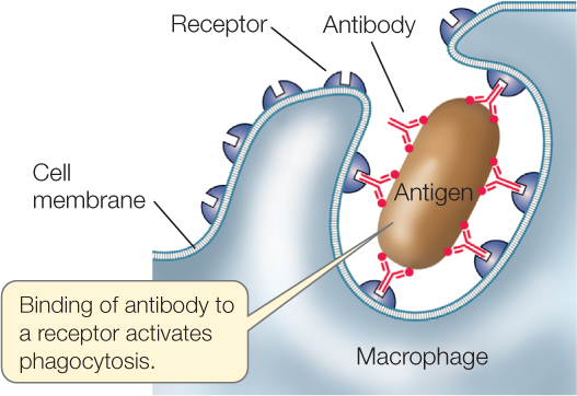
- Activation of the complement system: When antibodies bind to antigens that are present on the surfaces of pathogens, they attract components of the complement system (see Concept 39.2). Some complement proteins attack and lyse the pathogenic cells. In addition, the binding of antibody and complement proteins stimulates phagocytes (monocytes, macrophages, or dendritic cells) to ingest the pathogens.

- Activation of effector cells: Because of its cross-linking function (see Concept 39.4), antibody binding can result in large, insoluble antibody–pathogen or antibody–antigen complexes. These attract effector cells such as phagocytes and natural killer cells.
CHECKpointCONCEPT39.4
- Sketch an IgG antibody, identifying the variable and constant regions, light and heavy chains, and antigen-binding sites.
- Assume that one IgG heavy chain is encoded by 1,500 base pairs of DNA, and that one IgG light chain is encoded by 600 base pairs of DNA. If each antibody were encoded by a different gene, how many base pairs of DNA would be needed for 10 million antibody genes? Is it possible for the human genome to contain all these genes? Explain your answer.
- The bacterium that causes diphtheria (see Figure 39.5) synthesizes a toxic protein. You have probably not been exposed to this bacterium or its toxin. At the present time, are you making B cells and antibodies that bind specifically to diphtheria toxin? Explain your answer.
- When an antigenic protein such as diphtheria toxin (see above) enters the bloodstream, clones of numerous B cells are produced. Explain.
The humoral immune response works in concert with the cellular immune response in adaptive immunity. Now let’s turn to a closer examination of the cellular response.