Protein Composition and Structure
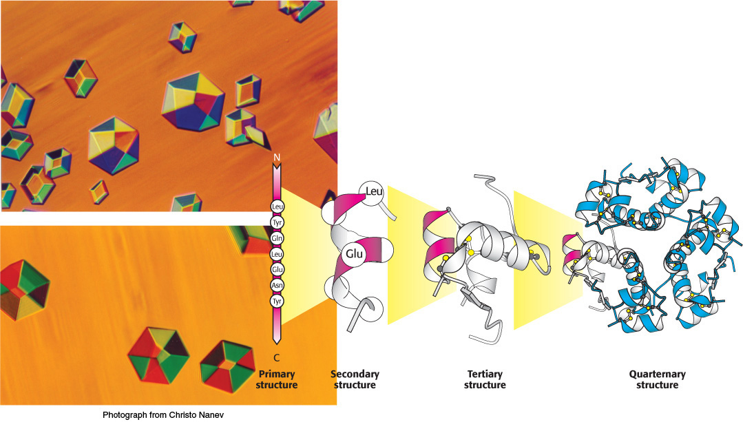
Insulin is a protein hormone, crucial for maintaining blood sugar at appropriate levels. (Below) Chains of amino acids in a specific sequence (the primary structure) define a protein such as insulin. Amino acids close to one another within this sequence can fold into regular structures (the secondary structure), such as the α-helix. Entire chains fold into well-defined structures (the tertiary structure)—in this case, a single insulin molecule. Such structures assemble with other chains to form arrays such as the complex of six insulin molecules shown at the far right (the quaternary structure). These arrays can often be induced to form well-defined crystals (photograph at left), which allows a determination of these structures in detail.
[Photograph from Christo Nanev.]
Proteins are the most versatile macromolecules in living systems and serve crucial functions in essentially all biological processes. They function as catalysts, transport and store other molecules such as oxygen, provide mechanical support and immune protection, generate movement, transmit nerve impulses, and control growth and differentiation. Indeed, much of this book will focus on understanding what proteins do and how they perform these functions.
Several key properties enable proteins to participate in a wide range of functions.
1. Proteins are linear polymers built of monomer units called amino acids, which are linked end to end. The sequence of linked amino acids is called the primary structure. Remarkably, proteins spontaneously fold up into three-dimensional structures that are determined by the sequence of amino acids in the protein polymer. Three-dimensional structure formed by hydrogen bonds between amino acids near one another is called secondary structure, whereas tertiary structure is formed by long-range interactions between amino acids. Protein function depends directly on this three-dimensional structure (Figure 2.1). Thus, proteins are the embodiment of the transition from the one-dimensional world of sequences to the three-dimensional world of molecules capable of diverse activities. Many proteins also display quaternary structure, in which the functional protein is composed of several distinct polypeptide chains.
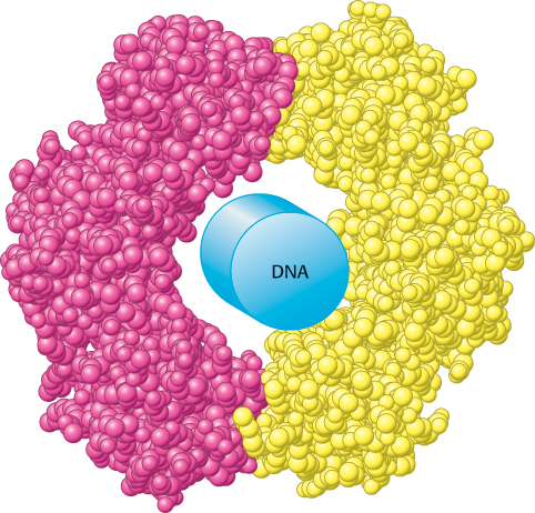
 FIGURE 2.1 Structure dictates function. A protein component of the DNA replication machinery surrounds a section of DNA double helix depicted as a cylinder. The protein, which consists of two identical subunits (shown in red and yellow), acts as a clamp that allows large segments of DNA to be copied without the replication machinery dissociating from the DNA.
[Drawn from 2POL.pdb.]
2. Proteins contain a wide range of functional groups. These functional groups include alcohols, thiols, thioethers, carboxylic acids, carboxamides, and a variety of basic groups. Most of these groups are chemically reactive. When combined in various sequences, this array of functional groups accounts for the broad spectrum of protein function. For instance, their reactive properties are essential to the function of enzymes, the proteins that catalyze specific chemical reactions in biological systems (Chapters 8 through 10).
3. Proteins can interact with one another and with other biological macromolecules to form complex assemblies. The proteins within these assemblies can act synergistically to generate capabilities that individual proteins may lack. Examples of these assemblies include macromolecular machines that replicate DNA, transmit signals within cells, and enable muscle cells to contract (Figure 2.2).
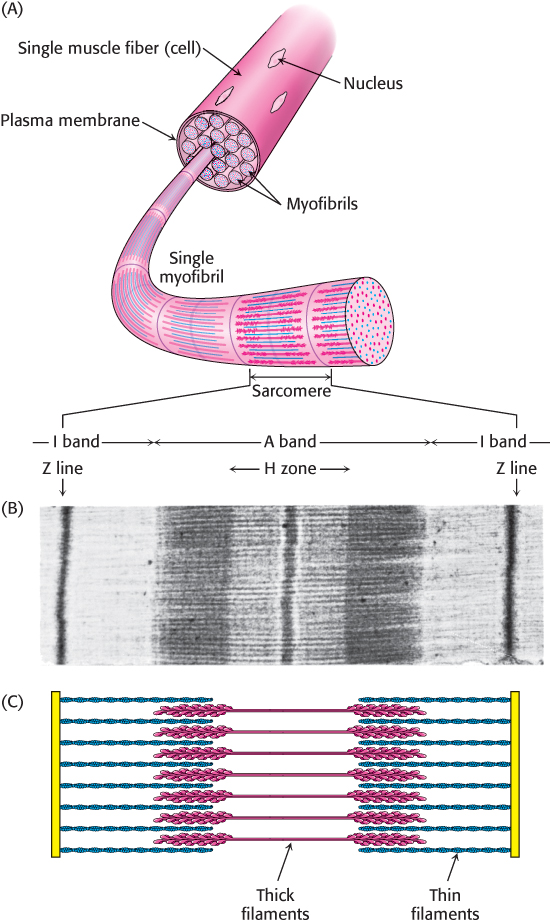
FIGURE 2.2A complex protein assembly. (A) A single muscle cell contains multiple myofibrils, each of which is comprised of numerous repeats of a complex protein assembly known as the sarcomere. (B) The banding pattern of a sarcomere, evident by electron microscopy, is caused by (C) the interdigitation of filaments made up of many individual proteins.
[(B) Courtesy of Dr. Hugh Huxley.]
4. Some proteins are quite rigid, whereas others display considerable flexibility. Rigid units can function as structural elements in the cytoskeleton (the internal scaffolding within cells) or in connective tissue. Proteins with some flexibility may act as hinges, springs, or levers. In addition, conformational changes within proteins enable the regulated assembly of larger protein complexes as well as the transmission of information within and between cells (Figure 2.3).
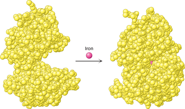
FIGURE 2.3 Flexibility and function. On binding iron, the protein lactoferrin undergoes a substantial change in conformation that allows other molecules to distinguish between the iron-free and the iron-bound forms.
[Drawn from 1LFH.pdb and 1LFG.pdb.]