13.2 ENZYMATIC MACHINES IN BACTERIAL RECOMBINATIONAL DNA REPAIR

The complex pathways shown in Figures 13-2 through 13-6 have been elucidated by genetic studies carried out in bacteria and eukaryotes. Many of the enzymes that promote the steps in these pathways have been characterized in vitro, and multiple steps of some pathways have been reconstituted with purified enzymes (Table 13-1). Complete reconstitution of a DSB repair or a replication fork repair remains a key goal of the field. In this section we describe the major enzyme systems found in bacteria, before moving on to eukaryotic recombination systems.

Figure 13-1: Enzymes/Proteins Involved in Bacterial Recombinational DNA Repair

RecBCD and RecFOR Initiate Recombinational Repair

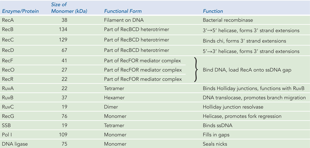
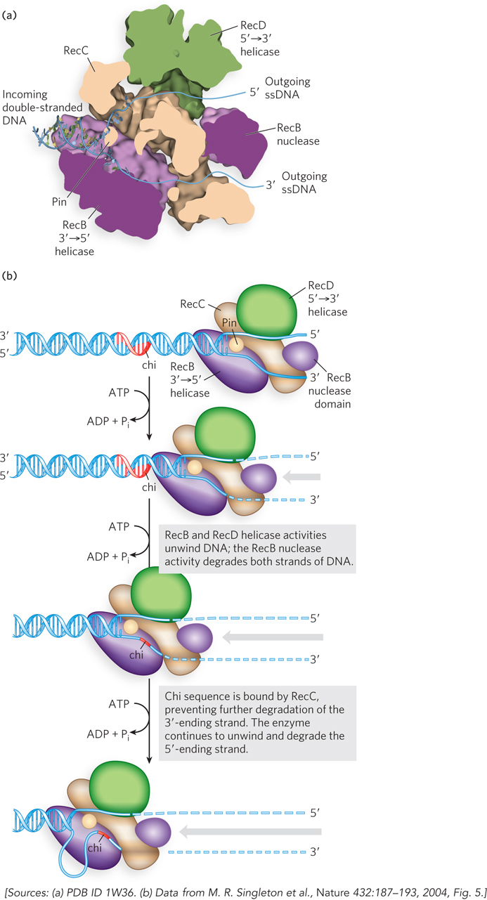
In all cells, the process of recombinational DNA repair revolves around a recombinase enzyme. In bacteria, that recombinase is the RecA protein. Before RecA can act, the stage must be set by other enzymes that degrade one strand of DNA, where necessary, and load RecA onto the DNA. The RecBCD and RecFOR complexes, with three subunits each, are the initiators of recombinational DNA repair. RecBCD loads the RecA protein onto DNA to repair double-strand breaks, and RecFOR does so to repair DNA gaps.

In E. coli, the recB, recC, and recD genes encode the heterotrimeric RecBCD enzyme, which has both helicase and nuclease activities. This enzyme has two jobs: (1) it prepares the 3′-ending single strand by degrading the 5′-ending strand at the site of a DSB, and (2) it directly loads the RecA protein onto the prepared single-stranded DNA tail. The RecBCD enzyme binds to linear DNA at a free (broken) end and moves inward along the double helix, unwinding and degrading the DNA in a reaction coupled to ATP hydrolysis. Initially, both DNA strands are degraded. The structure of RecBCD reveals a great deal about how the complex works (Figure 13-7a). The RecB and RecD subunits are helicase motors, with RecB moving in the 3′→5′ direction along one strand and RecD moving in the 5′→3′ direction along the other strand. One domain of the RecB subunit is also a nuclease that degrades both DNA strands as they are unwound. The activity of the enzyme is altered when it interacts with a sequence referred to as chi, 5′-GCTGGTGG-3′, on the 3′-ending strand, which binds tightly to a site on the RecC subunit. From that point, degradation of the strand with a 3′ terminus is greatly reduced, but degradation of the 5′-ending strand is increased (Figure 13-7b). This process creates the single-stranded 3′ overhang that initiates virtually all recombination events, as outlined in Figure 13-2. There are 1,009 chi sequences scattered throughout the E. coli genome, and these sites enhance the frequency of recombination about fivefold to tenfold in their immediate vicinity (within 1,000 bp). The chi octamer sequence is greatly overrepresented in the E. coli chromosome (relative to a random occurrence of a random eight-nucleotide sequence), and the chi sites are not randomly oriented in the DNA. Almost all have the orientation needed to facilitate the repair of DSBs that occur during replication.

Figure 13-7: The RecBCD helicase/nuclease. (a) The structure of the RecBCD enzyme. (b) Activities of the RecBCD enzyme at a DNA end. The RecB and RecD subunits travel along the DNA, a process that requires ATP; RecB degrades both strands as the complex travels, cleaving the 3′-ending strand more often than the 5′-ending strand. The RecC subunit has a protrusion called a pin that facilitates separation of the DNA strands. When a chi site is encountered on the 3′-ending strand, the RecC subunit binds to it, halting the advance of this strand through the complex. Degradation of the 5′-ending strand continues as the 3′-ending strand is looped out, eventually creating a 3′ single-stranded extension. RecA (not shown) is then loaded onto the processed DNA by the RecBCD enzyme.

458

As the 3′-ending strand is turned into a single strand, it is rapidly bound by the single-stranded DNA–binding protein (SSB) (see Figure 5-3). This protein directs traffic on segments of single-stranded DNA, blocking the access of some proteins and enzymes and facilitating the access of others. RecA is one of the proteins that is blocked, and SSB helps prevent the binding of RecA to single-strand gaps where recombination is not required (such as the transient gaps on the lagging strand during DNA synthesis). When recombinational DNA repair becomes necessary, RecA loading is mainly a process of overcoming this SSB barrier.

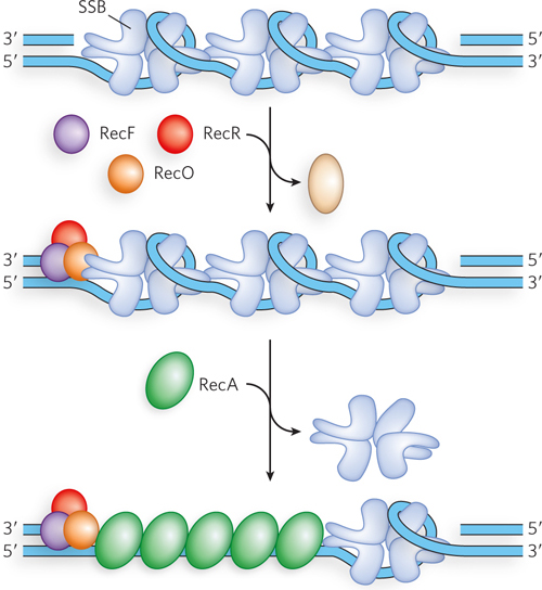
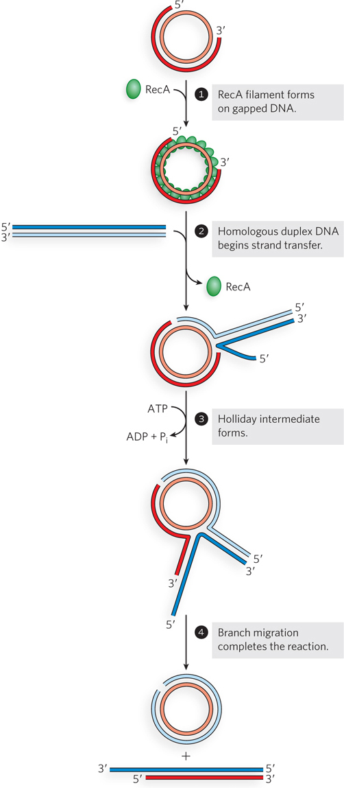
As we will see, the RecA protein forms a nucleoprotein filament on the DNA. The filament is formed in two steps: first the binding of a few RecA subunits (called nucleation), then extension of the filament from that initial complex. The nucleation step limits the overall process and is the step that is blocked by SSB. As the RecBCD enzyme unwinds the DNA and degrades the 5′-ending strand, a domain within the RecB subunit recruits RecA to the 3′-ending strand. This loading function of RecB serves to nucleate the RecA binding that is needed to promote strand invasion.

The RecA protein must also be loaded onto single-strand DNA gaps, such as the one shown in Figure 13-6 (top). These gaps are also bound by SSB, blocking access by RecA. In this case, the key loading factors are the RecF, RecO, and RecR proteins, collectively called RecFOR. The RecF protein is recruited by an unknown mechanism to the double-stranded DNA immediately adjacent to the gap, on the free 5′ end in the discontinuous strand. With the aid of RecO and RecR, the RecA protein is activated to displace SSB and nucleate the formation of the RecA filament needed for the subsequent phases of repair (Figure 13-8). Proteins that have the unique function of loading other proteins onto DNA are called mediator proteins. The RecFOR proteins are recombination mediators in most bacteria.

Figure 13-8: The role of RecFOR in loading RecA protein onto SSB-coated single-stranded DNA. SSB is displaced, and RecA is loaded.

459

RecA Protein Is the Bacterial Recombinase

The bacterial RecA protein is the prototype of a class of proteins found in all organisms, from bacteria to humans (see the How We Know section at the end of this chapter). The RecA-class recombinases promote the central steps in recombination reactions. They align a single-stranded DNA with a homologous double-stranded DNA and promote a strand switch that pairs the single strand with its complement in the duplex, displacing the other duplex strand. This is the reaction that we have been referring to as strand invasion.

RecA-class recombinases are unusual among the proteins of DNA metabolism in that their active forms are ordered, helical filaments of up to several thousand subunits that assemble cooperatively (Figure 13-9). RecA binds most readily to single-stranded DNA. As noted above, the filament forms in two steps, first nucleating on the single-stranded DNA (the step that is blocked by SSB), then growing by the addition of subunits, primarily in the 5′→3′ direction. A polar filament is created, with growth occurring at one end and most dissociation occurring at the other, trailing 5′-proximal end. RecA is a DNA-dependent ATPase. ATP is hydrolyzed by subunits throughout the RecA filament; when hydrolysis occurs in the subunit at the trailing end, it often results in dissociation of that subunit. Once formed, the RecA nucleoprotein filament is ready to promote the DNA strand exchanges at the heart of recombination.

Figure 13-9: RecA protein filaments. RecA and other recombinases in this class function as filaments of nucleoprotein. (a) Segment of a RecA filament with four helical turns (24 RecA subunits). Notice the bound double-stranded DNA in the center. The core domain of RecA is structurally related to the domains in helicases. (b) Electron micrograph of a RecA filament bound to DNA. (c) Filament formation proceeds in discrete nucleation and extension steps. Extension occurs by adding RecA subunits so that the filament grows in the 5′→3′ direction. When disassembly occurs, subunits are subtracted from the trailing end.

460

The RecA filament can facilitate strand exchange with a variety of substrates in vitro. The single-stranded DNA may be linear or circular, or a single-strand gap within double-stranded DNA. As RecA promotes strand invasion of these single-stranded substrates into a double-stranded DNA, a variety of structures result (Figure 13-10a–c). When the exchange is initiated in a DNA gap, branch migration may move the process into the adjacent duplex, where the exchange then involves four strands (Figure 13-10d). The reactions promoted with linear single strands best mimic the strand invasion that initiates most recombination reactions as outlined in Figure 13-2, as well as the repair of DSBs created during replication (see Figure 13-4). A circular single strand, easily purified from certain bacterial viruses, has been a particularly convenient DNA substrate for monitoring RecA activity in vitro. The single-strand-gapped substrate provides a model for reactions occurring during gap repair.

Figure 13-10: Recombination-related reactions promoted by the RecA protein of E. coli. (a) Strand invasion involving a 3′ single-stranded extension to form a D-loop. Here, RecA is promoting the strand invasion reaction outlined in Figure 13-2. (b) A DNA strand exchange involving three DNA strands. The substrates shown here are often used in research because the products are readily separated and visualized by agarose gel electrophoresis. The initial step is a strand invasion of the single-stranded DNA into one end of the duplex. Once strand invasion occurs, RecA also promotes a unidirectional (5′→3′ relative to the single-stranded circle) branch migration that completes an exchange of strands between the two DNA substrates. (c) DNA strand exchange involving a single-strand gap. Reactions of this sort also begin with strand invasion of the single strand into the duplex and are often facilitated by topoisomerases. (d) Strand exchange involving a single-strand gap and four DNA strands. The reaction begins with a strand invasion of the DNA in the single-strand gap into another duplex DNA. RecA then promotes a directional branch migration that carries the reaction into the adjacent duplex.

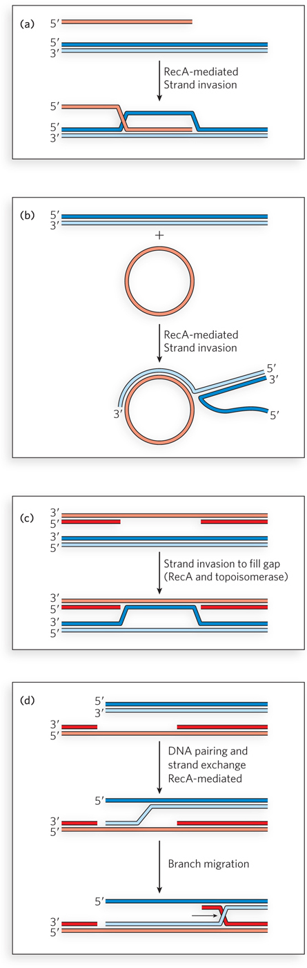
In each scenario, the single strand of DNA is first bound by RecA to establish the nucleoprotein filament. The RecA filament then promotes strand invasion into a homologous double-stranded DNA, aligning the bound single strand with the complementary strand in the duplex over a region that can involve hundreds of base pairs. The exchanged region can be further extended by RecA-promoted branch migration, in a process that requires ATP hydrolysis. The branch migration occurs at a rate of 6 bp/s at 37°C and progresses in the 5′→3′ direction relative to the single-stranded DNA in the RecA filament.

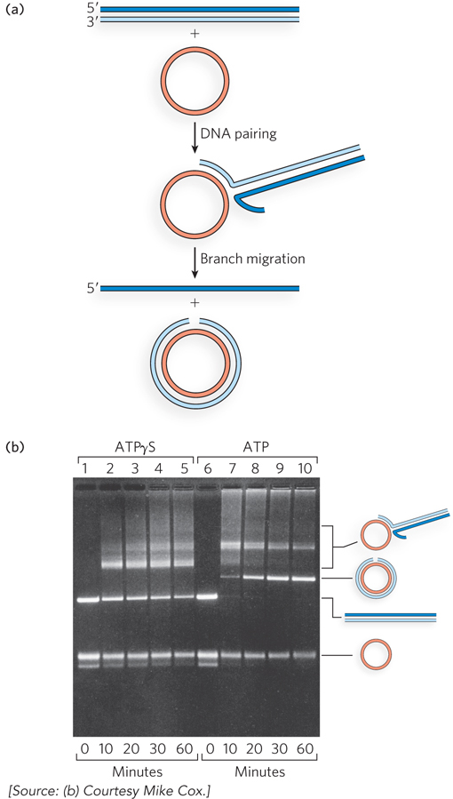
When purified RecA protein promotes DNA strand exchange in vitro between a circular single strand and a linear duplex, the substrates and products have distinctive structures that are readily separated and visualized by agarose gel electrophoresis (Figure 13-11). The initial pairing of the RecA-bound single-stranded circle and the linear duplex creates a branched DNA intermediate. After a period of RecA-promoted branch migration around the circle, a nicked circular duplex and a displaced linear single strand are formed as reaction products. The initial DNA alignment requires ATP, but not its hydrolysis. The facilitated branch migration necessary to complete exchange is coupled to ATP hydrolysis.

Figure 13-11: RecA-mediated DNA strand exchange. (a) The DNA substrates, intermediates, and products of the DNA strand exchange reaction. The circular DNAs, derived from bacteriophages, have a total length of 5,000 to 8,000 nucleotides or base pairs. With DNAs of this size, the reaction time is measured in tens of minutes at 37°C. (b) Intermediates (as diagrammed to the right of the agarose gel) generated by the initial strand invasion. Complete exchange of strands requires ATP hydrolysis. No branch migration occurs when ATPγS, a nonhydrolyzable ATP analog, replaces ATP (lanes 1 to 5).

461

If the circular substrate is a gapped double-stranded DNA (i.e., one strand is discontinuous), the DNA strand exchange is initiated in the single-strand gap, and RecA-promoted branch migration extends it into the double-stranded region (Figure 13-12). Because DNA is a helical structure, continued strand exchange requires an ordered rotation of the two aligned DNAs. The mechanism by which ATP hydrolysis is coupled to facilitated branch migration is not yet understood, but it probably entails the dissociation of RecA subunits at the trailing end of the RecA filament. When bound at a stalled replication fork, RecA can promote fork regression, using its capacity to promote branch migration.

Figure 13-12: Steps in a RecA-mediated DNA strand exchange reaction involving four DNA strands. Much RecA protein remains on the DNA throughout the reaction, although filament disassembly may be coupled to the movement of the branch point. The reaction begins with only three strands, initiated in the single-strand gap of one DNA substrate. As branch migration carries the branch into the duplex adjoining the gap, a Holliday intermediate forms.

RecA Protein Is Subject to Regulation

In principle, RecA-mediated recombination can occur between any two homologous DNA sequences. In every bacterial chromosome, some sequences, such as those that encode ribosomal RNAs, are repeated. Recombination between these sequences could have catastrophic consequences, leading to the deletion or rearrangement of large segments of the chromosome. For this reason, recombination, in general, and RecA protein activity, in particular, are highly regulated.

Regulation occurs at three levels: transcription of the recA gene, autoregulation, and regulation by other proteins. Transcriptional regulation occurs within the context of the bacterial SOS response (described in Chapter 20). Most regulation at other levels is directed at the formation, disassembly, and function of RecA protein filaments.

462

Autoregulation is “self” regulation. The RecA protein suppresses its own activities by means of a highly charged C-terminal peptide flap (see Figure 5-20). Removal of just 17 amino acid residues from the RecA C-terminus creates a RecA species for which almost all activities are enhanced. For example, whereas filament nucleation of native RecA protein is blocked by SSB, a C-terminal deletion mutant of RecA readily displaces SSB without the aid of RecBCD or RecFOR. In the mutant cell, this can result in elevated levels of recombination.

463

Many other proteins play a role in regulating the RecA protein. As we have seen, the RecBCD and RecFOR complexes facilitate the RecA filament nucleation process. Reliance on these loading functions helps direct RecA filament formation to DNA regions where it is needed. Another regulatory protein, RecX, binds to the growing RecA filament end and halts filament extension. A protein called DinI binds along the RecA filament and stabilizes it, while at the same time limiting the DNA strand exchange process. The helicase UvrD actively removes RecA filaments from the DNA when they are no longer needed. These and other proteins, working as an integrated system, help limit RecA function and direct it toward particular repair requirements.

Multiple Enzymes Process DNA Intermediates Created by RecA

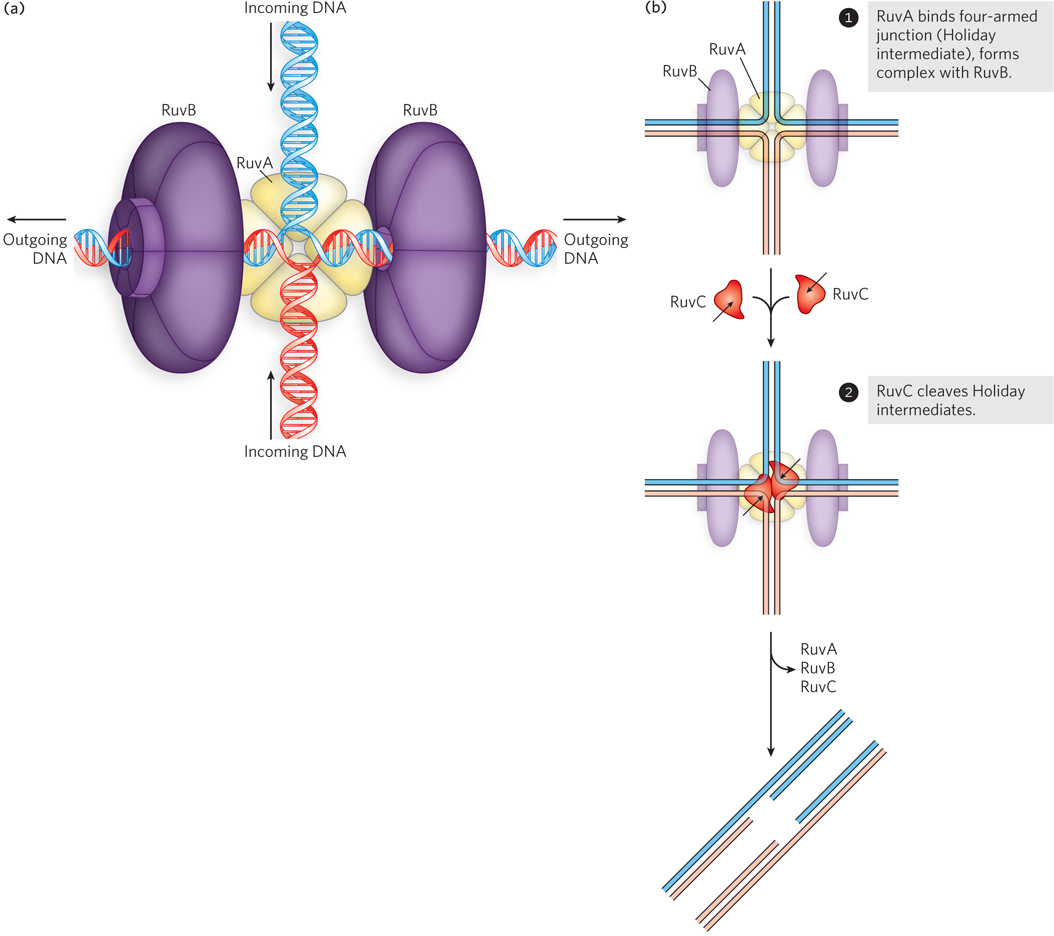
RecA is not the only protein in a bacterial cell that can promote branch migration; other enzyme systems are specialized for that task. As one example, the processing of Holliday intermediates is facilitated by a complex called RuvAB (repair of UV damage). Up to two RuvA protein tetramers bind to a Holliday intermediate and form a complex with two RuvB hexamers (Figure 13-13). The donut-shaped RuvB hexamers surround two of the four arms of the Holliday intermediate. RuvB is a DNA translocase, related in structure and function to hexameric DNA helicases. The DNA is propelled outward through the hole in the donut, away from the junction, in a reaction coupled to ATP hydrolysis. The result is very rapid branch migration that can move the position of the Holliday intermediate by thousands of base pairs in a few seconds. The RuvAB complex moves the Holliday intermediate away from the region of damaged DNA and recruits RuvC, a Holliday intermediate resolvase. RuvC replaces one of the RuvA tetramers at the junction and cleaves strands in opposing arms of the Holliday intermediate to resolve it into viable chromosomal products. The nicks in the DNA products are sealed by DNA ligase.

Figure 13-13: Catalysis of DNA branch migration and Holliday intermediate resolution by the RuvA, RuvB, and RuvC proteins. (a) RuvA binds to a four-armed junction (Holliday intermediate) and forms a complex with RuvB, a hexameric DNA translocase, on two sides. (b) RuvC cleaves Holliday intermediates (black arrows).

464

HIGHLIGHT 13-1 EVOLUTION:A Tough Organism in a Tough Environment: Deinococcus radiodurans

Some radioactive isotopes, such as 60Co and 137Cs, emit a type of ionizing radiation called γ rays. Gamma rays are photons; they transmit energy to atoms in solution, generating ions that include the highly reactive hydroxyl radical. In a living cell exposed to γ rays, any molecule can be damaged, including proteins and DNA. Double-strand breaks are included in the carnage.

The energy deposited by electromagnetic radiation is measured in rads or grays (1 Gy = 100 rads). For a human cell, a dose of 2 to 5 Gy is lethal. In the 1950s, it became clear that some organisms are surprisingly resistant to radiation. For example, in efforts to use radioactive sources to sterilize food, some sealed food samples were spoiled by bacterial action even after exposure to γ radiation at levels up to 4,000 Gy. The culprit was a pink, non-spore-forming, nonmotile bacterium eventually named Deinococcus radiodurans. D. radiodurans can absorb the damage inflicted by γ irradiation at 5,000 Gy with no lethality. A dose of this kind causes substantial damage even to a Pyrex beaker. More relevant to the cell, a 5,000 Gy dose produces many hundreds of DSBs, in addition to thousands of single-strand breaks and other lesions.

The Deinococcus genome consists of four circular DNA molecules, all generally present in multiple copies. After γ irradiation, the cells stop growing and DNA repair begins. Overlapping DNA fragments are spliced together, and the entire genome is accurately reconstituted within a few hours. The cells begin to grow and divide again as if nothing had happened. It is perhaps the most remarkable feat of DNA repair we know of so far.

This process is demonstrated in the gel shown in Figure 1. Following various treatments, D. radiodurans genomic DNA was isolated, treated with a restriction enzyme that cuts only a few times in the genome, and subjected to pulsed field gel electrophoresis (see Chapter 7). In cells grown under normal conditions, this procedure yields the series of large DNA fragments shown in the second lane of the gel (the first lane consists of markers of known molecular weight). Immediately after γ irradiation at 5,000 Gy, this banding pattern disappears, replaced by a smear of randomly sized, smaller DNA fragments. Over the next 3 to 4 hours, the normal band pattern reappears as the genome is accurately reconstituted.

FIGURE 1 DNA from γ-irradiated D. radiodurans is initially fragmented, but after several hours of repair it regains its normal banding pattern.

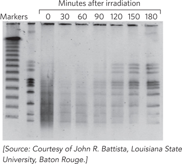
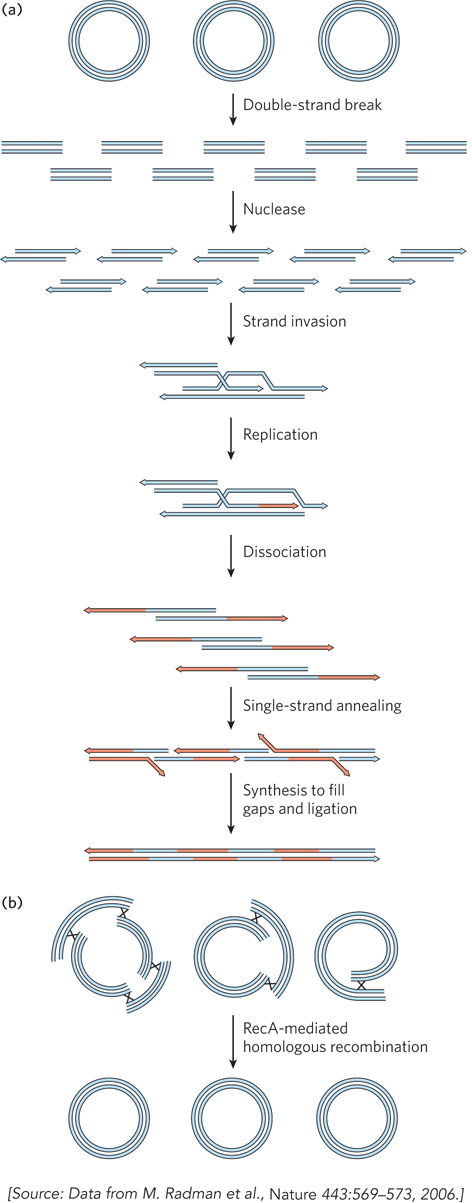
Genome reconstitution in D. radiodurans is recombinational DNA repair on a massive scale. The Deinococcus RecA protein (DrRecA) plays a key role, and most of the radiation resistance of this organism disappears if DrRecA is inactivated. There are two stages of repair, each requiring about 90 to 120 minutes under optimal conditions. The first stage uses a process similar to synthesis-dependent strand annealing (SDSA), but with an extended phase of replication (extended SDSA, or ESDSA). Once again, the process starts with the steps outlined in Figure 13-2. The ends of the broken DNA fragments are processed so as to degrade the 5′-ending strands, generating 3′ extensions (Figure 2a). These are then used in RecA-promoted strand invasion reactions. The 3′ ends act as primers for extended DNA synthesis, using a homologous chromosome strand as the template. After dissociation, the long 3′ extensions are annealed to each other where complementarity exists. The process is completed by nuclease treatment and ligation, as needed. The second stage again uses RecA to facilitate a larger-scale splicing of chromosomal segments (Figure 2b).

FIGURE 2 (a) The first stage of double-strand break repair in D. radiodurans closely resembles synthesis-dependent strand annealing (SDSA). (b) In the second stage of genomic reconstitution, large chromosomal segments are spliced together by DrRecA protein.

For several decades, D. radiodurans was considered the most radiation-resistant organism known. Recent research, however, has revealed many microbial species that are highly resistant to radiation, some of them more so than D. radiodurans. In addition, these highly resistant species are found in various unrelated genera, indicating that this phenotype has evolved independently many times. There are no environments on Earth that are subject to ionizing radiation at levels of thousands of grays, but these bacteria are not aliens from outer space. A few species have been found in hot springs with high radon backgrounds, where exposure to chronic low levels of radiation has forced adaptations to permit more efficient DNA repair. However, the most reliable source of bacteria with extreme radiation resistance is a desert environment, where the major selective pressure is not radiation but desiccation. When water disappears for an extended period of time, most metabolic processes, including generation of the ATP needed for DNA repair, cease. However, spontaneous DNA damage continues, and DSBs are among the accumulating lesions. The extreme radiation resistance of D. radiodurans and many other bacteria reflects their extraordinary capacity for rapid genomic reconstitution when desiccation gives way to conditions favorable for growth.

465

The bacterial recombination systems that repair replication forks can also repair DSBs created by ionizing radiation, sometimes with startling proficiency. The bacterium Deinococcus radiodurans can survive and prosper after absorbing doses of ionizing radiation sufficient to generate thousands of DSBs (Highlight 13-1).

The repair of stalled or collapsed replication forks is generally followed by a restart of replication. A five-protein complex called the restart primosome loads the replicative helicase, DnaB, onto the DNA at the reconstituted replication fork. The rest of the replisome assembles around DnaB, and replication starts anew.

466

Repair of the Replication Fork in Bacteria Can Lead to Dimeric Chromosomes

Some pathways of replication fork repair lead to the creation of a Holliday intermediate behind the reconstituted fork instead of in front of it (Figure 13-14; see also Figure 13-5). This Holliday intermediate can be resolved by RuvC in one of two ways: by cleaving the crossover strands (shown as resolution path X in Figure 13-14) or by cleaving the template strands (resolution path Y). The first path, cleaving the crossover strands, simply leads to the completion of replication and the segregation of two monomeric chromosomes into daughter cells. The second, cleaving the template strands, has a special consequence when the genome is circular, as it is for most bacterial chromosomes: it ultimately creates a single dimeric chromosome that cannot be segregated at cell division. Under normal growth conditions, this outcome is observed in about 15% of cells in an E. coli culture.

Figure 13-14: The generation and resolution of dimeric chromosomes formed during bacterial replication. There are two ways to resolve any Holliday intermediate. In path X, the crossover strands are cut and ligated to form two separate chromosomes. In path Y, the crossover strands are cut and ligated to form a contiguous, dimeric form of the circular chromosome, when replication is complete.

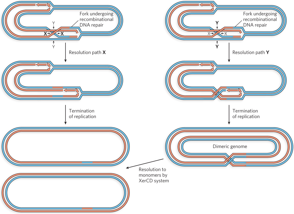
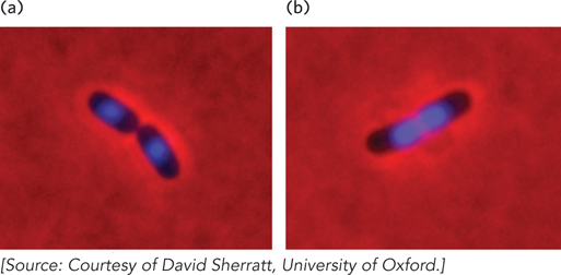
Cells harboring dimeric chromosomes do not die. Instead, the stalled chromosomal segregation is detected, triggering the activity of a specialized site-specific recombination system, called XerCD, that converts the dimer back into monomeric circles (see Figure 13-14). Site-specific recombination is a class of reaction discussed in Chapter 14. Once the monomeric chromosomes are generated, cell division completes normally. If the cells have a mutation that inactivates the site-specific recombination system, cells with dimeric chromosomes become “stuck,” unable to divide (Figure 13-15). For these cells, the mutation is lethal.

Figure 13-15: Cell division is hindered in bacterial cells lacking the capacity to resolve dimeric chromosomes. (a) Wild-type E. coli cells immediately after cell division. (b) E. coli cells with a mutation that inactivates the dimeric chromosome resolution system, after chromosomal division. In both photos, the chromosomes were condensed by treatment with chloramphenicol and stained with a blue fluorescent dye.

SECTION 13.2 SUMMARY

  • The bacterial RecBCD and RecFOR complexes provide pathways for loading the RecA protein onto single-stranded extensions (at double-strand breaks) or onto single-strand gaps, respectively.

  • The bacterial RecA protein is the prototypical recombinase, forming a filament on single-stranded DNA and promoting strand invasion reactions.

  • Recombination is a highly regulated process, and the RecA protein is the major target for regulation, which occurs through transcriptional regulation, autoregulation, and regulation by other proteins.

    467

  • Recombination intermediates generated by RecA are processed by enzymes such as the RuvA, RuvB, and RuvC proteins.

  • In the circular bacterial chromosome, resolution of Holliday intermediates associated with replication fork repair can lead to the formation of dimeric chromosomes.