CONCEPT32.1 Circulatory Systems Can Be Closed or Open
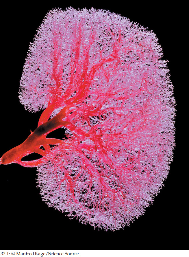
A circulatory system typically consists of a muscular pump (heart), a fluid (blood), and a series of conduits (blood vessels or channels) through which the blood is pumped. Because blood must flow close to all the cells of the body, the profusion of blood vessels is often spectacular when visualized (FIGURE 32.1).
For an animal to have a high degree of division of labor among its organs, a circulatory system is essential because it allows each organ to export its products promptly to other parts of the body and to receive promptly the products of other organs. With a circulatory system, O2 taken up by gills or lungs can be delivered rapidly to all other organs (see Figure 31.3); hormones secreted by endocrine glands can be distributed quickly throughout the body; lipid molecules stored in body fat can be delivered rapidly to the cells of any organ where they are needed; and waste products of cell metabolism can be carried promptly to the kidneys for excretion.
The sophistication of the circulatory system in a species tends to be correlated with the species’ need for circulatory O2 transport. This is because O2 transport is typically the most urgent circulatory function. Many minutes can go by without any transport of hormones, food molecules, or wastes—animals can tolerate such interruptions. However, animals typically cannot tolerate long interruptions in O2 transport. For instance, if a person’s blood circulation stops during a cardiac arrest, depriving brain cells of O2, the person becomes unconscious within 20 seconds! When we consider how rapidly blood must flow in a circulatory system, the need for O2 transport is typically the most important consideration because—of all the substances carried by blood flow—O2 must be transported the fastest. This explains why animals that have evolved high metabolic rates (and therefore have high O2 needs) also usually have evolved high-performance circulatory systems.
The vertebrate animals nicely illustrate the relationship between metabolic rate and circulatory function. Mammals and birds typically have far higher rates of blood flow and higher blood pressures than lizards, frogs, or fish of the same body size. This difference in circulatory performance correlates with differences in metabolic rates. Mammals and birds have far higher metabolic rates than lizards, frogs, or fish (see Concept 29.3) and therefore require circulatory systems that can deliver O2 far faster. The same relationship is seen among types of fish. Fish species with high metabolic rates—such as the tuna in our opening story—have circulatory systems capable of higher performance than those of sluggish fish species. Similarly, among the mollusks, squid—which are fast-swimming predators—have high-performance circulatory systems compared with those of sedentary mollusks such as snails and clams.
Insects are the exception that proves the rule. Some insects, when flying, have very high O2 needs per gram of body weight. Yet insects have modest, low-performance circulatory systems. The tracheal breathing system of insects accounts for this paradox. O2 is brought to each cell of an insect by a branch of the animal’s breathing system, not by blood circulation. The circulatory system, freed of responsibility to transport O2, need not perform at a high level, despite the high metabolic rates of insects.
LINK
The breathing system of insects is described in Concept 31.2; see especially Figure 31.10
Closed circulatory systems move blood through blood vessels
A circulatory system is closed if the blood is always contained within blood vessels as it circulates. The vertebrates again provide excellent examples and will be our focus here. The heart of a vertebrate pumps blood into large blood vessels called arteries (which we will discuss in more detail in Concept 32.2). The arteries branch like trees into smaller and smaller blood vessels as they carry the blood to tissues and organs throughout the body. The branches ultimately become so small in diameter that we cannot see them with the naked eye. Nonetheless, the blood always remains within vessels. As blood leaves tissues and organs, it flows through vessels that merge into larger and larger vessels—the veins—that carry it back to the heart:
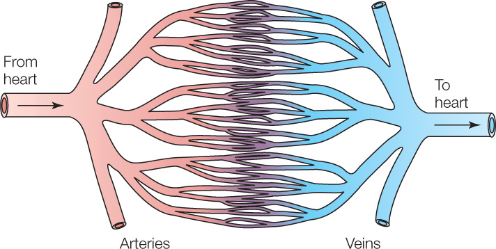
The blood vessels of smallest diameter—which we can see only with a microscope—are collectively called the vessels of the microcirculation and consist of arterioles, capillaries, and venules. The arterioles and capillaries are particularly critical for the functioning of a closed circulatory system. As we discuss them, an important point to recognize is that in the walls of all blood vessels, the layer of cells that immediately surrounds the blood is a simple epithelium (see Figure 29.13). For historical reasons, this layer of cells is called the vascular endothelium.
The walls of capillaries consist only of vascular endothelium. In other words, they consist only of a single layer of thin, flattened epithelial cells, less than 1 μm thick (Figure 32.2A). Moreover, capillaries are of tiny diameter. The open passage in a blood vessel is called the vessel lumen. In capillaries, the lumen is often just slightly larger in diameter than individual red blood cells. For this reason, the cells (∼7–8 μm in diameter in humans) must pass through capillaries in single file (Figure 32.2B). In each tissue of the body, capillaries are found near every tissue cell. For instance, capillaries are positioned near every brain cell and every muscle cell. The number of capillaries is so great as to be almost unimaginable. Added together, for example, the capillaries in a single cubic centimeter of mammalian skeletal muscle or heart muscle are 10–30 meters long!
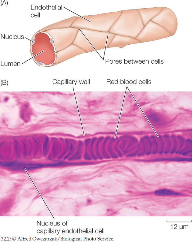
Capillaries are the principal sites in a closed circulatory system where blood releases O2, nutrient molecules, hormones, and other substances to the tissues. They are also the principal sites where blood takes up CO2 and other waste products from the tissues. Capillaries play these roles in part because they are so numerous. They are also suited to these roles because their walls are thin and often porous, allowing materials to move rapidly across their walls by diffusion and other means.
Arterioles are as important for the function of the microcirculation as capillaries are. The walls of an arteriole consist of an inner layer of vascular endothelium surrounded by an outer layer of smooth muscle cells that are under the control of the autonomic nervous system (described in Concept 34.5) and other control mechanisms. The muscle cells in the wall can adjust the diameter of an arteriole’s lumen. Contraction of the muscle cells causes vasoconstriction, reducing the lumen diameter. Relaxation of the muscle cells allows the lumen diameter to increase, a process called vasodilation. Control of the arteriole lumen diameter in these ways is called vasomotor control.
Why do changes in the lumen diameter matter? They are of enormous importance because they control where blood flows. The famous French physiologist Jean Poiseuille (pronounced Pwa-sul) demonstrated in the mid-19th century that the rate of fluid flow through a tube is extraordinarily sensitive to the diameter of the tube lumen because this rate depends directly on the 4th power of the diameter. In other words, if the diameter of the lumen of a tube is doubled, the flow rate through the tube increases by a factor of 16! Conversely, halving the diameter reduces flow to 1/16 of its previous rate. An arteriole precedes each capillary bed (set of interconnected capillaries) and controls the flow of blood into it (FIGURE 32.3). By controlling the diameters of the arterioles, the autonomic nervous system and other systems exert control over which capillary beds receive blood flow.
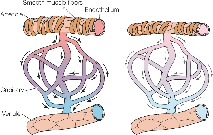
Several everyday examples illustrate arteriolar control of blood flow. The skin of people with fair complexions is often more pinkish in warm than in cold weather. This change reflects vasomotor control. Skin arterioles dilate in warm weather, permitting vigorous blood flow to the capillary beds near the skin surface—a response that makes the skin turn pink and helps metabolic heat leave the body. When people exercise, arterioles in their exercising skeletal muscles dilate, and arterioles in many other parts of their body constrict. These changes increase blood flow to the exercising muscles. Erection of the penis is one of the most familiar cases of arteriolar control. During sexual arousal, dilation occurs in arterioles that supply blood to the spongy tissue of the penis, permitting the spongy tissue to inflate with blood under high pressure (see Concept 37.2).
In general, closed circulatory systems can support higher levels of metabolic activity than the open systems we will discuss next, in part because they have a higher capacity to direct blood flow to the tissues that need it the most. In addition to vertebrates, other animals with closed circulatory systems are the annelid worms, such as earthworms, and the cephalopod mollusks, such as squid and octopuses.
In open circulatory systems, blood leaves blood vessels
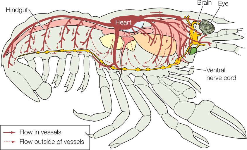
A circulatory system is open if the blood exits blood vessels as it flows through the body. Arthropods—such as insects, spiders, crabs, and lobsters—have open circulatory systems, as do most mollusks. In an animal with an open circulatory system, the heart typically pumps blood into arteries that carry the blood for at least a short distance. Ultimately, however, the vessels end, and the blood pours out into spaces surrounded not by vascular endothelium but by ordinary tissue cells. After this, the blood makes its way back to the heart, not in veins but by flowing through channels called sinuses and lacunae. The sinuses and lacunae are (respectively) small and large spaces among ordinary tissue cells.
In an open circulatory system, no distinction exists between blood (the fluid pumped by the heart and flowing in the blood vessels) and interstitial fluid (the fluid found between cells in the ordinary tissues—also called tissue fluid; see Concept 29.4). Debate exists about the correct name to use for the fluid in open circulatory systems. Here we call it blood. Some biologists, however, prefer to call the fluid hemolymph, referring to the fact that it has attributes of both blood (hemo, “blood”) and tissue fluid (lymph). Tissue cells are directly bathed by the blood or hemolymph. For this reason, the cells can readily take up O2 and other materials from the blood, or release wastes into it.
Open circulatory systems are not very well understood, although we know they sometimes have very high rates of blood flow. They are thought to be limited in how well they can control where the blood goes. Circulation in a crayfish or lobster illustrates the pattern of flow in an open circulatory system (FIGURE 32.4). The heart pumps blood into arteries that carry the blood to most major parts of the body before ending. After the blood leaves the vessels, it makes its way through sinuses and lacunae to ventral blood spaces along the bases of the walking legs, where the gills arise. Sucking forces developed by the heart then pull the blood through the gills and back into the heart to be pumped again into the arteries.
Circulatory systems can be closed or open
Atherosclerosis (“hardening of the arteries”) is a potentially deadly chronic disease. The immediate problem is that fatty deposits—composed of lipids such as cholesterol and triacylglycerols—build up in blood vessels. Take out a ruler, and using the principles developed by Poiseuille (principles known today as Poiseuille’s Law), estimate how much more slowly blood will pass through the atherosclerotic artery at the right, compared with the healthy artery to the left, assuming everything is the same except the fatty deposits. People start their lives with arteries like the one at the left.

CHECKpointCONCEPT32.1
- What is the most fundamental distinction between open and closed circulatory systems?
- How do arterioles and capillaries differ structurally and functionally?
- A grasshopper and a clam both have open circulatory systems. What enables the grasshopper to be more active and sustain a higher metabolic rate?
For the rest of this chapter, we will focus mainly on the closed circulatory systems of vertebrates. One reason for this focus is that we ourselves have a closed circulatory system. We’ll start by discussing the major patterns of blood flow throughout the vertebrate body.