15-4 Cerebral Asymmetry in Thinking
Seizing on Sperry’s findings, the 1980s saw an avalanche of self-
Fundamental to behavioral neuroscience was the finding by Paul Broca and his contemporaries in the mid-
Anatomical Asymmetry
Building on Broca’s findings, investigators have learned how the language-
Figure 15-11 shows that the lateral fissure, which partly separates the temporal and parietal lobes, has a sharper upward course in the right hemisphere relative to the left. As a result, the posterior right temporal lobe is larger than the same region on the left, as is the left parietal lobe relative to the right.
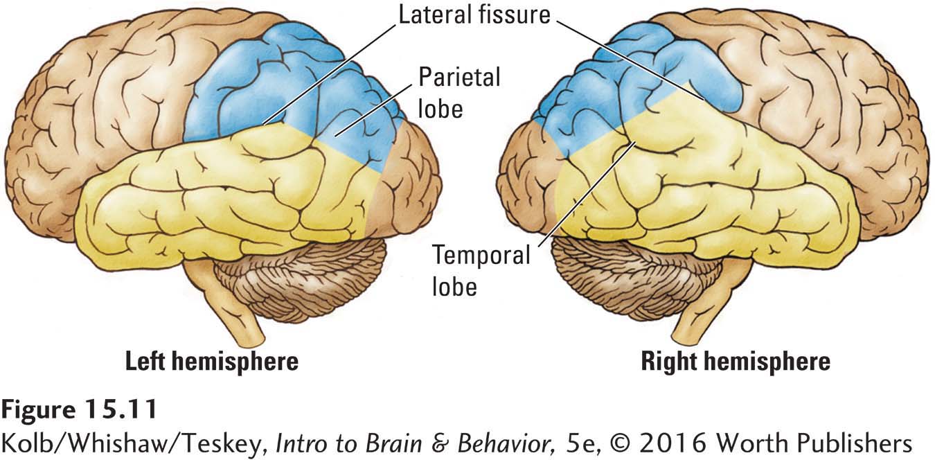
Among the anatomical asymmetries in the frontal lobes, the region of sensorimotor cortex representing the face is larger in the left hemisphere than in the right, a difference that presumably corresponds to the left hemisphere’s special role in talking. Broca’s area is organized differently on the left and the right. The area visible on the brain’s surface is about one-
Not only do these gross anatomical differences exist but so too do hemispheric differences in the details of their cellular and neurochemical structures. For example, neurons in Broca’s area on the left have larger dendritic fields than do corresponding neurons on the right. The discovery of structural asymmetries told us little about the reasons for such differences, but ongoing research is revealing that they result from underlying differences in cognitive processing by the brain’s two sides.
Although many anatomical asymmetries in the human brain are related to language, brain asymmetries are not unique to humans. Most if not all mammals have asymmetries, as do many bird species. The functions of cerebral asymmetry therefore cannot be limited to language processing. Rather, human language likely evolved after the brain became asymmetrical. Language simply took advantage of processes, including the development of mirror neurons, that had already been lateralized by natural selection in earlier members of the human lineage.
Functional Asymmetry in Neurological Patients
The specialized functions of the cerebral hemispheres become obvious in people with damage to the left or right side of the brain. To see these functional differences clearly, compare the cases of G. H. and M. M.
Right Parietal Damage
When G. H. was 5 years old, as he was hiking with his family, a large rock rolled off an embankment and hit him on the head. He was unconscious for a few minutes and had a severe headache for a few days but quickly recovered. Around age 18, however, he started having seizures. Neurosurgical investigation revealed that G. H. had had a right posterior parietal injury from the rock accident. Figure 15-12A shows the area affected. After surgery to remove this area, G. H. had weakness on the left side of his body and showed contralateral neglect. But these symptoms lessened fairly quickly, and a month after the surgery, they had completely cleared.
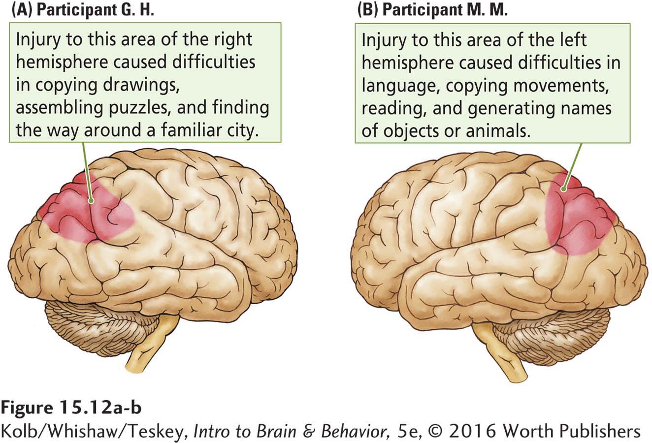
Nevertheless, G. H. had chronic difficulties in copying drawings; 4 years later, he still performed this task at about the level of a 6-
Left Parietal Damage
Meningioma is imaged in Focus 3-3.
M. M.’s difficulties were quite different. A meningioma had placed considerable pressure on the left parietal region. The tumor was surgically removed when M. M. was 16 years old. It had damaged the area shown in Figure 15-12B.
Section 10-4 describes how left-
After the surgery, M. M. had various problems, including aphasia, impaired language use. The condition lessened over time: a year after the surgery, M. M. spoke fluently. Unfortunately, other difficulties persisted. In solving arithmetic problems, in reading, and even in simply calling objects or animals by name, M. M. performed at about a 6-
Lessons from Patients G. H. and M. M
What can we learn about brain function by comparing G. H. and M. M.? Their lesions were in approximately the same location but in opposite hemispheres, and their symptoms were very different.
Judging from G. H.’s difficulties, the right hemisphere contributes to controlling spatial skills, such as drawing, assembling puzzles, and navigating in space. In contrast, M. M.’s condition reveals that the left hemisphere seems to contribute to controlling language functions and cognitive tasks related to schoolwork—
To some extent, then, the left and right hemispheres think about different types of information. The question is whether these functional differences can be observed in a healthy brain.
Functional Asymmetry in the Healthy Brain
In the course of studying the auditory capacity of people with a temporal lobe lesion, Doreen Kimura (1967) found something unexpected. She presented her control participants with two strings of digits, one played into each ear, a procedure known as dichotic listening. The task was to recall as many digits as possible.
Kimura found that the controls recalled more digits presented to the right ear than to the left. This result is surprising because the auditory system crosses repeatedly, beginning in the midbrain. Nonetheless, information coming from the right ear seems to have preferential access to the left (speaking) hemisphere.
In a later study, Kimura (1973) played two pieces of music for participants, one to each ear. She then gave the participants a multiple-
The demonstration of this functional asymmetry in the healthy brain provoked much interest in the 1970s, leading to demonstrations of functional asymmetries in the visual and tactile systems as well. Consider the visual system. If we fixate on a target, such as a dot positioned straight ahead, all the information to the left of the dot goes to the right hemisphere and all the information to the right of the dot goes to the left hemisphere, as shown in Figure 15-14.
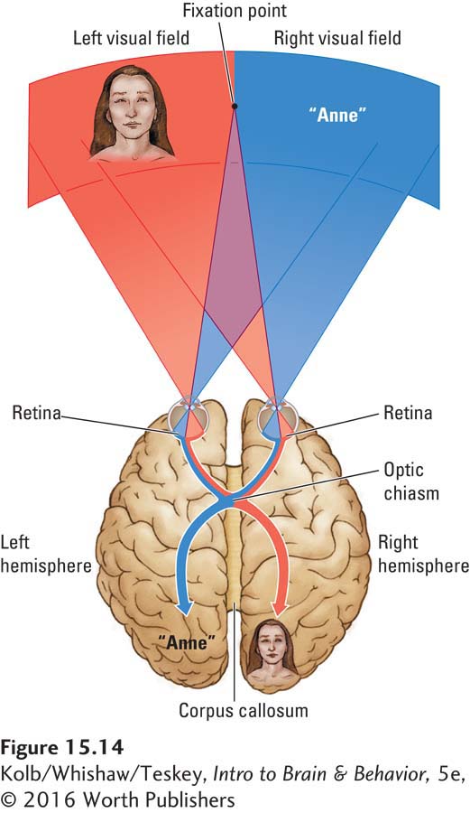
If information is presented for a relatively long time—
Words presented briefly to the right visual field and hence sent to the left hemisphere are more easily reported than are words presented briefly to the left visual field. Similarly, if complex geometric patterns or faces are shown briefly, those presented to the left visual field and hence sent to the right hemisphere are more accurately reported than are those presented to the right visual field.
Apparently, the hemispheres not only think about different types of information, they also process information differently. The left hemisphere seems biased toward processing language-
Those popular left brain–
A word of caution: Although asymmetry studies are fascinating, what they tell us about the differences between the hemispheres is not entirely clear. They tell us something is different, but it is a broad leap to conclude that the hemispheres house entirely different skill sets.
The hemispheres have many common functions, such as controlling movement in the contralateral hand and processing sensory information through the thalamus. Still, differences in the hemispheres’ cognitive operations do exist. We can better understand these differences by studying split-
Functional Asymmetry in the Split Brain
Before considering the details of split-
But how does a severed corpus callosum affect how the brain thinks? After the corpus callosum has been cut, the hemispheres have no way of communicating with one another. The left and right hemispheres are therefore free to think about different things. In a sense, a split-
One way to test the hemispheres’ cognitive functions in a split-
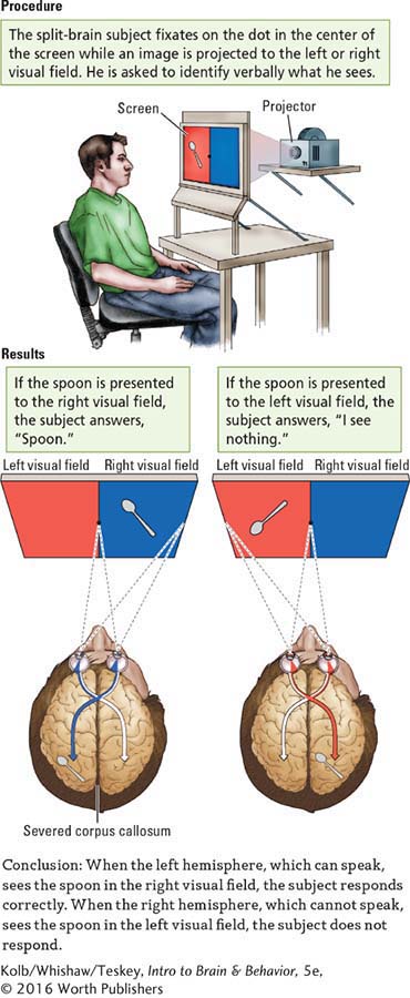
Experiments 15-3 and 15-4 show some basic testing procedures based on this dichotomy. The split-
EXPERIMENT
Question: Will severing the corpus callosum affect the way in which the brain responds?
EXPERIMENT
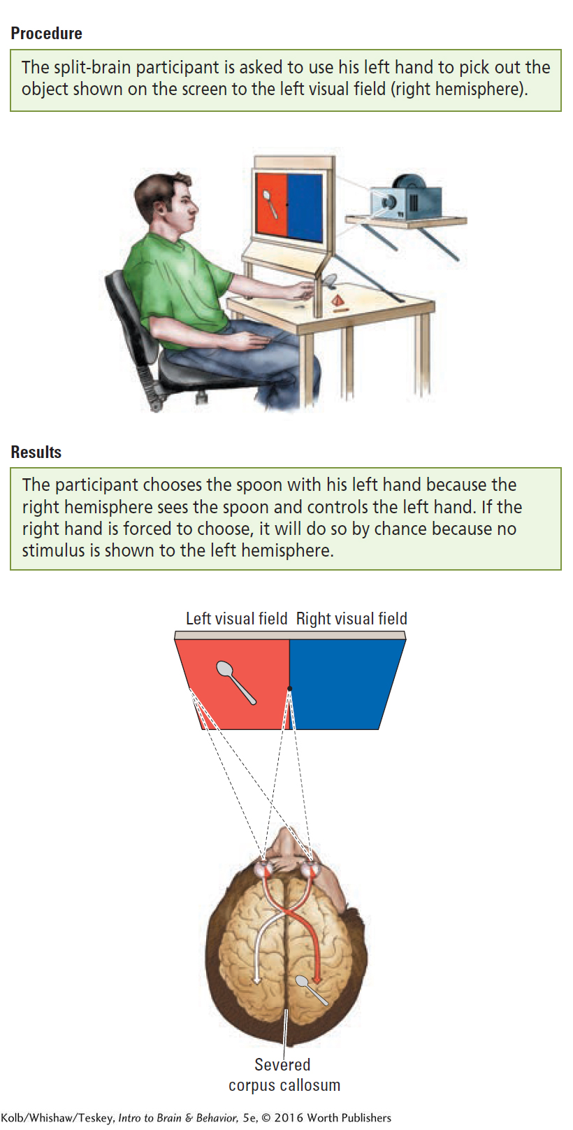
(A) Question: How can the right hemisphere of a split-
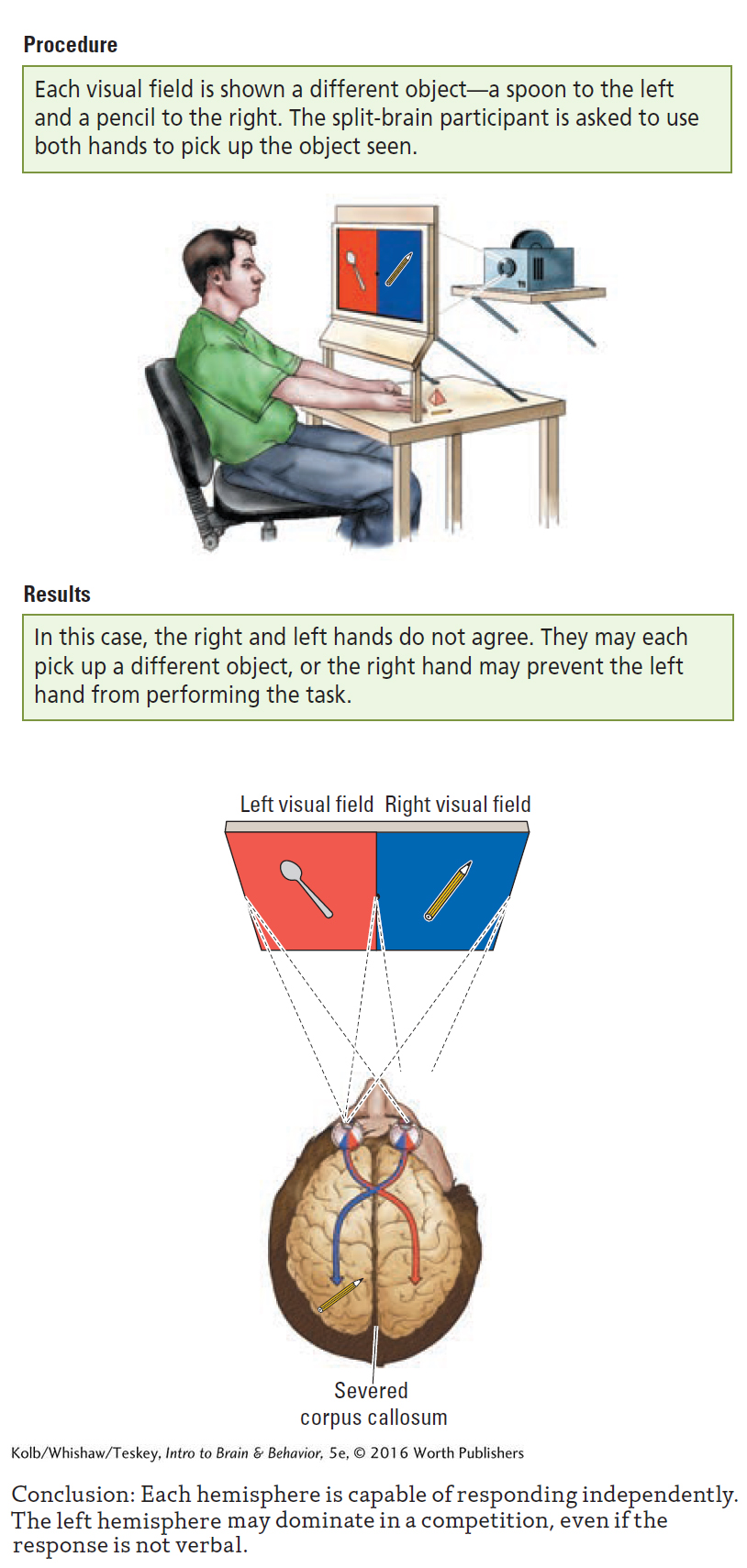
(B) Question: What happens if both hemispheres are asked to respond to competing information?
As illustrated in Experiment 15-3, for instance, a picture—
The right hemisphere (which receives the visual input) does not talk, so it cannot respond verbally, even though it sees the spoon in the left visual field.
The left hemisphere does talk, but it does not see the spoon, so it answers—
quite correctly, from its own perspective— that nothing was presented.
Now suppose the task changes. In Experiment 15-4A, the picture of a spoon is still presented to the left visual field, but the subject is asked to use the left hand to pick out the object shown on the screen. In this case, the left hand, controlled by the right hemisphere, which sees the spoon, readily picks out the correct object. Can the right hand also choose correctly? No, because it is controlled by the left hemisphere, which cannot see the spoon. If the person in this situation is forced to select an object with the right hand, the left hemisphere does so at random.
Now let’s consider an interesting twist. In the Procedure for Experiment 15-4B, each hemisphere is shown a different object—
This conflict between the hemispheres can be seen in the everyday behavior of some split-
We know from Experiment 15-3 that the left hemisphere is capable of using language, and Research Focus 15-1 reveals that the right hemisphere has visuospatial capabilities that the left hemisphere does not. Although findings from half a century of studying split-
Explaining Cerebral Asymmetry
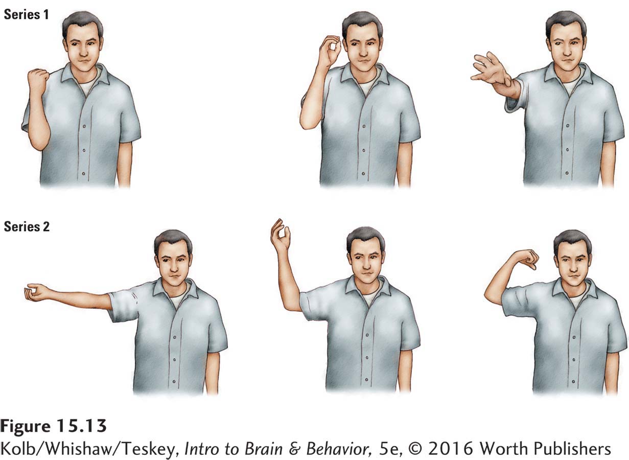
Various hypotheses propose to explain hemispheric differences. One idea, that the left hemisphere is important in controlling fine movements, dates back a century. Recall M. M., the meningioma patient with left parietal lobe damage and apraxia (see Figure 15-12B). Although the apraxia subsided, she was left with chronic trouble in copying movements.
Perhaps one reason that the left hemisphere has a role in language is that speaking requires fine motor movements of the mouth and tongue. Significantly, damage to the language-
That said, another clue that the left hemisphere’s specialization for language may be related to its special role in controlling fine movements comes from investigating where the brain processes certain parts of speech. Recall that cognitive systems for representing abstract concepts likely are related to systems that produce more concrete behaviors. Consequently, we might expect that the left hemisphere would participate in forming concepts related to fine movements.
Concepts that describe movements are the parts of speech we call verbs. A fundamental difference between left-
If the left hemisphere excels at language because it is better at controlling fine movements, what is the basis of the right hemisphere’s abilities? One idea is that the right hemisphere specializes in controlling movements in space. In a sense, this role elaborates the functions of the dorsal visual stream (diagrammed in Figure 15-3).
Once again, we can propose a link between movement at a concrete level and movement at a more abstract level. If the right hemisphere is producing movements in space, then it is also likely to produce mental images of such movements. We would therefore predict impairment of right-
The nervous system produces movement within a perceptual world the brain constructs.
Bear in mind that theories about the reasons for hemispheric asymmetry are highly speculative. The brain has evolved to produce movement and to construct a sensory reality, so the observed asymmetry must somehow relate to these overriding functions. That is, more recently evolved functions, such as language, likely are extensions of preexisting functions. So the fact that language is represented asymmetrically does not mean that the brain is asymmetrical because of language. After all, other species that do not talk have an asymmetrical brain.
Left Hemisphere, Language, and Thought
As we end our examination of brain asymmetry, consider one more provocative idea. Michael Gazzaniga (1992) proposed that the left hemisphere’s superior language skills are important for understanding the differences between humans’ and other animals’ thinking. He called the speaking hemisphere the interpreter. The following experiment, using split-
Each hemisphere is shown a picture of a match followed by a picture of a piece of wood, for example. Another set of pictures is then shown. The task is to pick from this set a third picture that has an inferred relation to the first two. In this example, the third related picture might be a bonfire. The right hemisphere is incapable of making the inference that a match struck and held to a piece of wood could start a bonfire, whereas the left hemisphere easily arrives at this interpretation.
An analogous task uses words. One or the other hemisphere might be shown the words pin and finger then be asked to pick out a third word related to the other two. In this case, the correct answer might be bleed.
The right hemisphere cannot make this connection. Although it has enough language ability to pick out close synonyms for pin and finger (needle and thumb, respectively), it cannot make the inference that pricking a finger with a pin will result in bleeding.
Again, the left hemisphere has no difficulty with this task. Apparently, the left hemisphere’s language capability gives it a capacity for interpretation that the right hemisphere lacks. One reason may be that language serves to label and express the computations of other cognitive systems.
Gazzaniga goes even further. He suggests that the evolution of left-
Gazzaniga’s idea is intriguing. It implies a fundamental difference in the nature of cerebral asymmetry—
15-4 REVIEW
Cerebral Asymmetry in Thinking
Before you continue, check your understanding.
Question 1
The right hemisphere plays a role in ____________ and ____________.
Question 2
The left hemisphere plays a role in ____________ and ____________.
Question 3
The split brain results from cutting apart the ____________.
Question 4
Why does it matter that the two cerebral hemispheres process information differently?
Answers appear in the Self Test section of the book.