2-4 Somatic Nervous System: Transmitting Information
The SNS is monitored and controlled by the CNS—
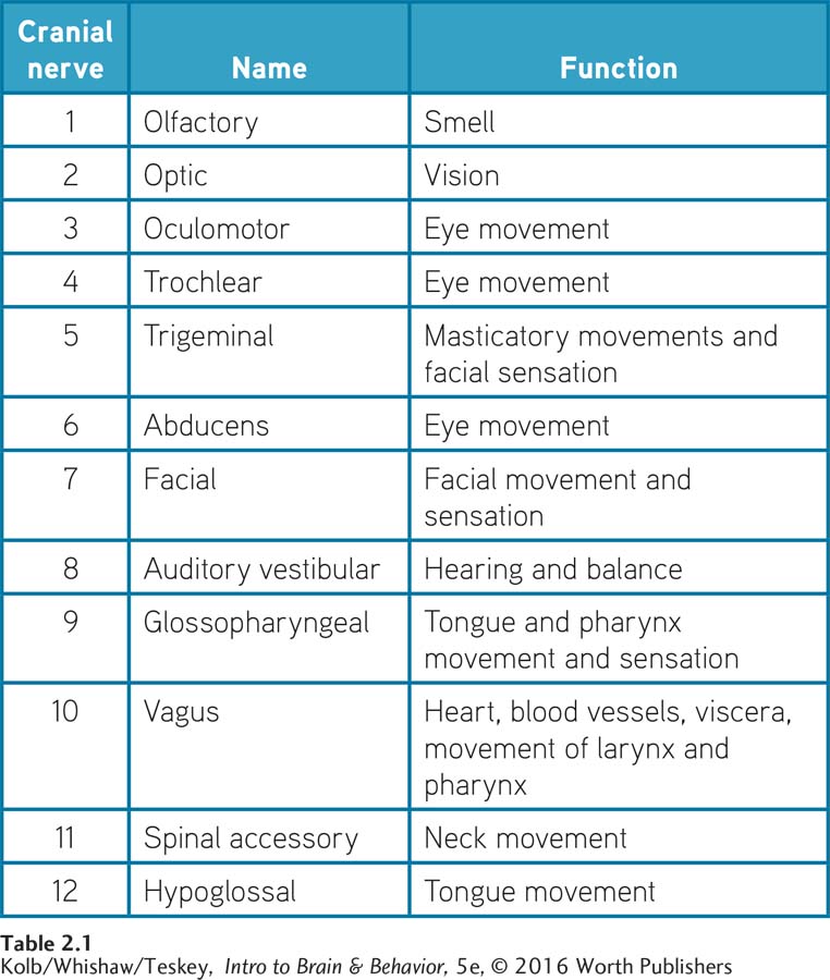
Cranial Nerves
The linkages provided by the cranial nerves between the brain and various parts of the head and neck as well as various internal organs are illustrated and tabulated in Figure 2-27. Cranial nerves can have afferent functions, such as sensory inputs to the brain from the eyes, ears, mouth, and nose, or they can have efferent functions, such as motor control of the facial muscles, tongue, and eyes. Some cranial nerves have both sensory and motor functions, such as modulation of both sensation and movement in the face.
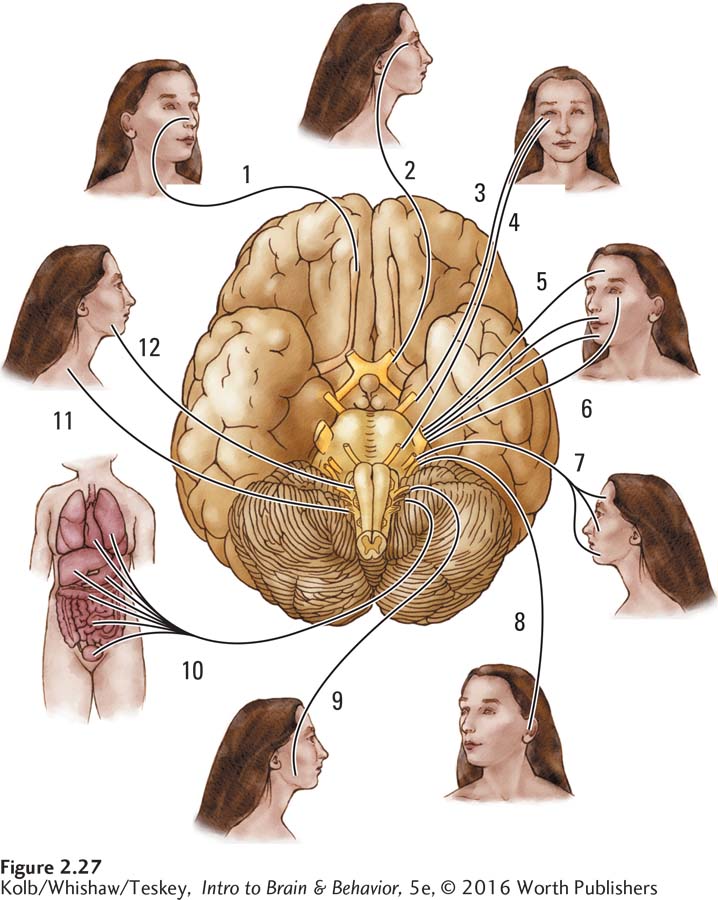
The 12 pairs of cranial nerves are known both by their numbers and by their names, as listed in Figure 2-27. One set of 12 controls the left side of the head, whereas the other set controls the right side. This arrangement makes sense for innervating duplicated parts of the head (such as the eyes), but why separate nerves should control the right and left sides of a singular structure (such as the tongue) is not so clear. Yet that is how the cranial nerves work. If you have ever received lidocaine (often called Novocaine) for dental work, you know that usually just one side of your tongue becomes numb because the dentist injects the drug into only one side of your mouth. The rest of the skin and muscles on each side of the head are similarly controlled by cranial nerves located on the same side.
We consider many cranial nerves in detail in later chapters’ discussions on topics such as vision, hearing, olfaction, taste, and stress responses. For now, you simply need to know that cranial nerves form part of the SNS, providing inputs to the brain from the head’s sensory organs and muscles and controlling head and facial movements. Some cranial nerves also contribute to maintaining autonomic functions by connecting the brain and internal organs (the vagus, cranial nerve 10) and by influencing other autonomic responses, such as salivation.
Spinal Nerves
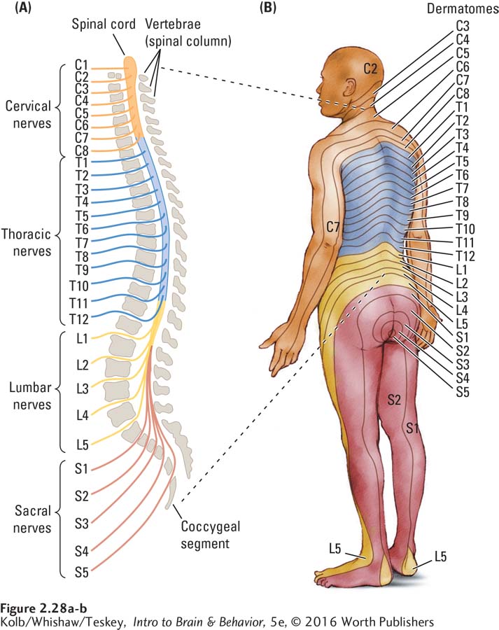
The spinal cord lies inside the bony spinal column, which is made up of a series of small bones called vertebrae (sing. vertebra), categorized into five anatomical regions from top to bottom: cervical, thoracic, lumbar, sacral, and coccygeal, as diagrammed in Figure 2-28A. You can think of each vertebra in these five groups as a short segment of the spinal column. The corresponding spinal cord segment in each vertebral region functions as that segment’s minibrain.
This arrangement may seem a bit odd, but it has a long evolutionary history. Think of a simpler animal, such as a snake, that evolved long before humans did. A snake’s body is a segmented tube. In that tube is another tube, the spinal cord, which also is segmented. Each of the snake’s nervous system segments receives nerve fibers from sensory receptors in the part of the body adjacent to it, and that nervous system segment sends fibers back to the muscles in that body part. Each segment, therefore, works independently.
Sections 11-1 and 11-4 review the spinal cord’s contributions to movement and to somatosensation.
A complication arises in animals such as humans, whose limbs may originate at one spinal segment level but extend past other segments of the spinal column. Your shoulders, for example, may begin at C5 (cervical segment 5), but your arms hang down well past the sacral segments. So unlike the snake, which has spinal cord segments that connect to body segments fairly directly adjacent to them, human body’s segments fall schematically into more of a patchwork pattern, as shown in Figure 2-28B. This arrangement makes sense if the arms are extended as they are when we walk on all fours.
Regardless of their complex pattern, however, our body segments still correspond to the spinal cord segments. Each of these body segments is called a dermatome (meaning skin cut). A dermatome has both a sensory nerve to send information from the skin, joints, and muscles to the spinal cord and a motor nerve to control the muscle movements in that particular body segment.
These sensory and motor nerves, known as spinal (or peripheral) nerves, are functionally equivalent to the cranial nerves of the head. Whereas the cranial nerves receive information from sensory receptors in the eyes, ears, facial skin, and so forth, the spinal nerves receive information from sensory receptors in the rest of the body—
Somatic Nervous System Connections
Like the CNS, the SNS is bilateral (two-
Figure 2-29A shows the spinal column in cross section. Look first at the nerve fibers entering its posterior side. These posterior fibers (dorsal in four-
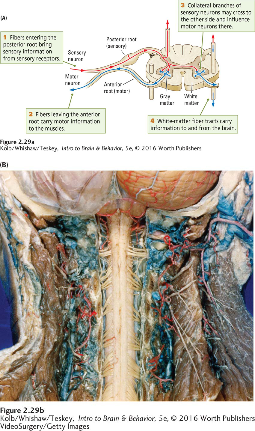
Fibers leaving the spinal cord’s anterior side are efferent, carrying information out from the spinal cord to the muscles. They, too, bundle together as they exit the spinal cord and so form an anterior root (ventral root in four-
Sections 11-1 and 11-4 explore spinal cord injuries and treatments, and Section 12-3, the link between spinal injury and loss of emotion.
The observation that the dorsal spinal cord is sensory and the ventral side is motor in four-
Integrating Spinal Functions
So far we have emphasized the spinal cord’s segmental organization, but the spinal cord must also somehow coordinate inputs and outputs across different segments. For example, many body movements require coordinating muscles controlled by different segments, just as many sensory experiences require coordinating sensory inputs to different parts of the spinal cord. How is this coordination accomplished? The answer is that the spinal cord segments are interconnected in such a way that adjacent segments can operate together to direct rather complex coordinated movements.
Integrating spinal cord activities does not require the brain’s participation, which is why the headless chicken can run around. Still, a close working relation must exist between the brain and the spinal cord. Otherwise, how could we consciously plan and execute our voluntary actions?
Somehow information must be relayed back and forth, and examples of this information sharing are numerous. For instance, tactile information from sensory nerves in the skin travels not just to the spinal cord but also to the cerebral cortex through the thalamus. Similarly, the cerebral cortex and other brain structures can control movements through their connections to the spinal cord’s anterior roots. So even though the brain and spinal cord can function independently, the two are intimately connected in their CNS functions.
2-4
Magendie, Bell, and Bell Palsy
François Magendie, a volatile and committed French experimental physiologist, reported in a three-
In 1811, a Scot named Charles Bell proposed functions for these nerve roots on the basis of anatomical information and the results of somewhat inconclusive experiments on rabbits. Although Bell’s findings were not identical with those of Magendie, they were similar enough to ignite a controversy. Bell hotly disputed Magendie’s claim to the discovery of dorsal and ventral root functions. As a result, the principle of sensory and motor segregation in the nervous system has been named after both researchers: the law of Bell and Magendie.
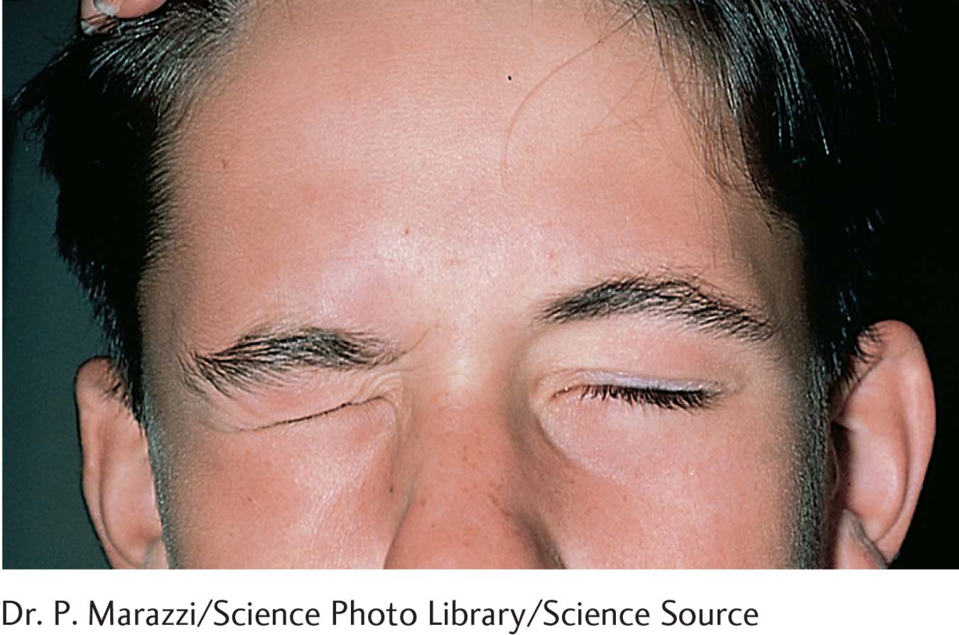
Magendie’s conclusive experiment on puppies was considered extremely important because for the first time it enabled neurologists to localize nervous system damage from the symptoms that a patient displays. Bell went on to describe an example of such localized cranial motor nerve dysfunction, and it still bears his name—
The onset of Bell palsy is typically sudden. Often the stricken person wakes up in the morning and is shocked to discover the face paralyzed on one side. He or she cannot open the mouth or completely close the eye on that side of the head. Most people fully recover from Bell palsy, although it may take several months. But in rare instances, such as that of Jean Chrétien, a former prime minister of Canada, partial paralysis of the mouth is permanent.
2-4 REVIEW
Somatic Nervous System: Transmitting Information
Before you continue, check your understanding.
Question 1
Two sets of SNS nerves, the __________ and the __________, receive sensory information or send motor signals to muscles or both.
Question 2
Both sets of SNS nerves are symmetrically organized, and each set controls functions on the __________ side of the body.
Question 3
The cranial nerves have both sensory and motor functions, receiving and sending information to the __________ and to the __________.
Question 4
Define the law of Bell and Magendie and explain why it is important.
Answers appear in the Self Test section of the book.