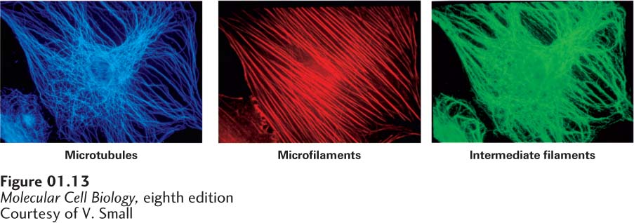
The cytoplasm contains an array of fibrous proteins collectively called the cytoskeleton (see Chapters 17 and 18). Three classes of fibers compose the cytoskeleton: microtubules (20 nm in diameter), built of polymers of the protein tubulin; microfilaments (7 nm in diameter), built of the protein actin; and intermediate filaments (10 nm in diameter). All of these fibers are long chains of multiple copies of one or more small protein subunits (Figure 1-13). The cytoskeleton gives the cell strength and rigidity, thereby helping to maintain its shape; this is perhaps most obvious with neurons, in which microtubules and other fibers allow the formation of the long, slim protuberances—the axons and dendrites (see Figure 1-3e and Chapter 22)—that emanate from the cell body and allow each neuron to carry out its specialized functions. Cytoskeletal fibers also control movement of structures within the cell; for example, some cytoskeletal fibers connect to organelles or provide tracks along which organelles and chromosomes move. Other fibers play key roles in cell motility. Perhaps most important, cell division and the segregation of chromosomes and organelles into the two daughter cells could not occur without the organizational framework provided by the cytoskeleton and its associated proteins.
[Courtesy of V. Small.]
FIGURE 1-13The three types of cytoskeletal filaments have characteristic distributions within mammalian cells. Three views of the same cell. A cultured fibroblast was permeabilized and then treated with three different antibody preparations. Each antibody binds specifically to the protein monomers forming one type of filament and is chemically linked to a differently colored fluorescent dye (green, blue, or red). Visualization of the stained cell in a fluorescence microscope reveals the locations of filaments bound to a particular dye-antibody preparation. In this case, microtubules are stained blue; microfilaments, red; and intermediate filaments, green. All three fiber systems contribute to the shape and movements of cells.
[Courtesy of V. Small.]
[NIBSC/Science Source.]
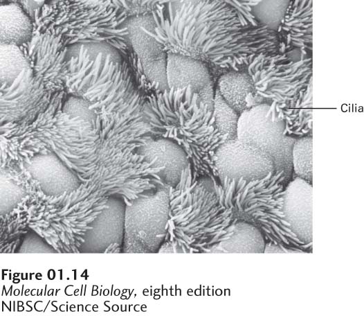
FIGURE 1-14Surface of the ciliated epithelium lining a mammalian trachea viewed in a scanning electron microscope. Beating cilia, which have a core of microtubules, propel mucus and foreign particles out of the respiratory tract, keeping the lungs and airways clear.
[NIBSC/Science Source.]
Cilia and flagella are similar extensions of the plasma membrane. They contain a bundle of microtubules that gives them shape and, together with motor proteins, allows them to beat rhythmically. They propel materials across epithelial surfaces (Figure 1-14), enable sperm to swim, and push eggs through the oviduct (see Chapter 18). As detailed in Chapter 16, most vertebrate cells contain at least one cilium that plays a key role in cell-cell signaling.