Growth of Cells in Two-Dimensional and Three-Dimensional Culture Mimics the In Vivo Environment
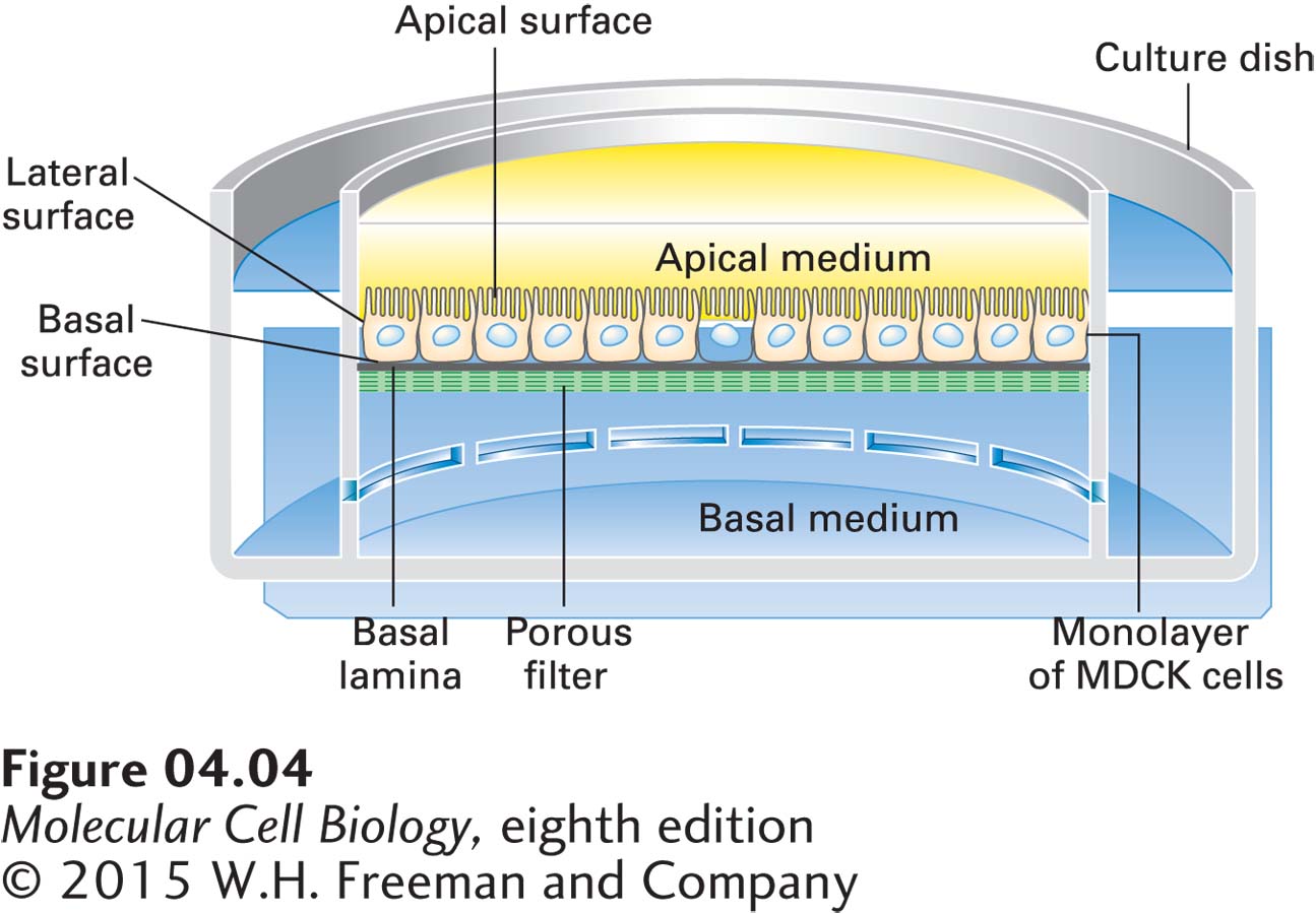
While much has been learned using cells grown on plastic or glass surfaces, these surfaces are far removed from cells’ normal tissue environment. As detailed in Chapter 20, many cell types function only when closely linked to other cells. Key examples are the sheet-like layers of epithelial tissue, called epithelia (singular, epithelium), that cover the external and internal surfaces of organs. Typically, epithelial cells have distinct surfaces, called the apical (top), basal (base or bottom), and lateral (side) surfaces (see Figure 20-11). The basal surface usually contacts an underlying extracellular matrix called the basal lamina, whose composition and function are discussed in Section 20.3. Epithelial cells often function to transport specific classes of molecules across the epithelial sheet; for example, the epithelial lining of the intestine transports nutrients into the cell through the apical surface and out toward the bloodstream across the basolateral surface. When grown on plastic or glass, epithelial cells cannot easily perform this function. Therefore, special containers have been designed with a porous surface that acts as a basal lamina to which epithelial cells attach and form a uniform two-dimensional sheet (Figure 4-4). A commonly used cultured cell line derived from dog kidney epithelium, called the Madin-Darby canine kidney (MDCK) cell line, is often used to study the formation and function of epithelial sheets.
FIGURE 4-4Madin-Darby canine kidney (MDCK) cells grown in specialized containers provide a useful experimental system for studying epithelial cells. MDCK cells form a polarized epithelium when grown on a porous membrane filter coated on one side with collagen and other components of the basal lamina. With the use of the special culture dish shown here, the medium on each side of the filter (apical and basal sides of the monolayer) can be experimentally manipulated and the movement of molecules across the layer monitored. Several cell junctions that interconnect the cells form only if the growth medium contains sufficient calcium.
Page 134
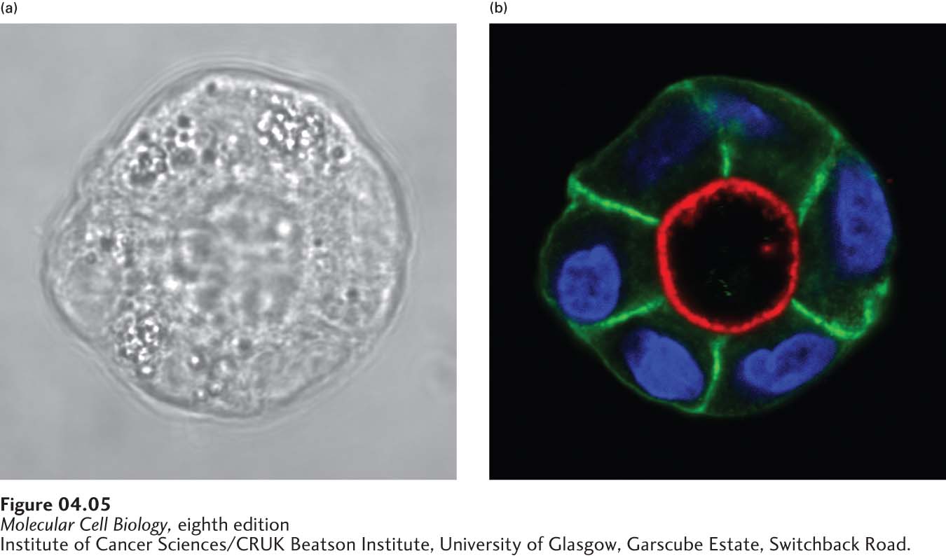
However, even a two-dimensional sheet often does not allow cells to fully mimic behavior in their normal environment. Methods have now been developed to grow cells in three dimensions by providing a support infiltrated with appropriate components of the extracellular matrix (discussed in Chapter 20). If MDCK cells are cultured under appropriate conditions, they will form a tubular sheet mimicking a tubular organ or the duct of a secretory gland. In these three-dimensional structures, the apical side of the epithelial sheet lines the lumen, whereas the basal side of each cell is in contact with the extracellular matrix (Figure 4-5).
[Institute of Cancer Sciences/CRUK Beatson Institute, University of Glasgow, Garscube Estate, Switchback Road.]
EXPERIMENTAL FIGURE 4-5MDCK cells can form cysts in culture. (a) MDCK cells grown on a supported extracellular matrix will form groups of cells that polarize to form a tubular single layer of cells with a lumen in the middle, called a cyst. (b) By examining the localization of proteins found in the apical (red) and basolateral membranes (green), we can see that these cells are fully polarized, with the apical side facing the lumen, which recapitulates their organization in the kidney tubules from which they are derived. The nuclear DNA is stained blue.
[Institute of Cancer Sciences/CRUK Beatson Institute, University of Glasgow, Garscube Estate, Switchback Road.]
Page 135
If we can grow an epithelial tube in culture, can we grow a whole organ that could be transplanted into a patient? Recent advances in biomedical engineering are developing promising strategies to do this, initially in experimental animals. In an example of one approach, a 3-D printer is used to help make a replacement ear. First, an exact computer image of an ear is generated. This image is used to program a 3-D printer to assemble a pliable matrix—containing support material that is biodegradable, together with appropriate components of the extracellular matrix—in the precise shape of an ear. This matrix provides support for the growth of skin cells, either in culture or after transplantation under the skin, so that ultimately the synthetic organ can be surgically attached to a living animal. Other approaches make use of 3-D printers to assemble the matrix and seed it with appropriate cells. An exciting and ambitious goal of this technology is to generate synthetic organs containing many different types of cells by printing each of several layers with appropriate matrix and cells to generate complex three-dimensional organs that might one day be used to replace defective ones in patients. Many hurdles still need to be overcome, but the ability to generate stem cells from patients and then induce differentiation in culture (described in Chapter 21) is overcoming the major obstacle of immunological rejection and will probably be key to providing cells for the assembly of synthetic organs.