14-5 Recovery from Brain Injury
The nervous system appears conservative in its use of mechanisms related to behavioral change. If neuroscientists wish to change the brain, as after injury or disease, then they should look for treatments that will produce plastic changes related to learning, memory, and other behaviors.
Recall that H. M. failed to recover his lost memory capacities, even after 55 years of practice in trying to remember information. Relearning simply was not possible for H. M. He had lost the requisite neural structures. But other people do show some recovery.
An average person would probably say that the recovery process after brain trauma requires the injured person to relearn lost skills, whether walking, talking, or using the fingers. But what exactly does recovery entail? Partial recovery of function is common after brain injury, but a person with brain trauma or brain disease has lost neurons. The brain may be missing structures critical for relearning or remembering.
Donna’s Experience with Traumatic Brain Injury
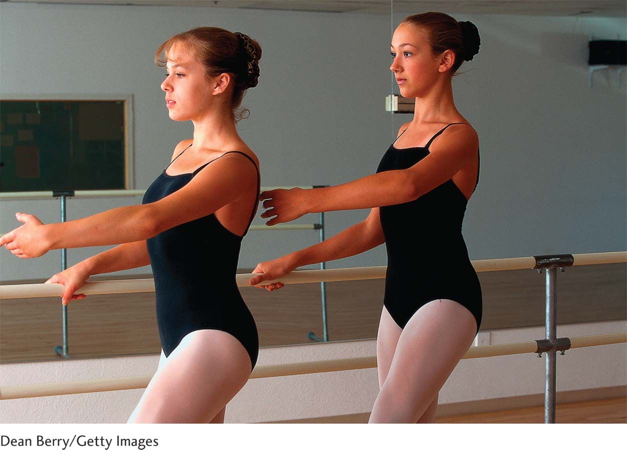
Donna started dancing when she was 4 years old, and she was a natural. By the time she finished high school, she had the training and skill necessary to apprentice with and later join a major dance company. Donna remembers vividly the day she was chosen to dance a leading role in The Nutcracker. She had marveled at the costumes as she watched the popular Christmas ballet as a child, and now she would dance in those costumes!
The births of two children interrupted her career as a dancer, but Donna never lost the interest. In 1968, when both her children were in school, she began dancing again with a local company. To her amazement, she could still perform most of the movements, although she was rusty on the choreography of the classical dances that she had once memorized so meticulously. Nonetheless, she quickly relearned. In retrospect, she should not have been so surprised, because she had always had an excellent memory.
Focus 1-1 and Section 1-2 introduce consequences of and treatments for TBI, which we elaborate here and in Section 16-3.
One evening in 1990, while on a bicycle ride, a drunk driver struck Donna. She was wearing a helmet but received a brain-
Over the ensuing 10 months, Donna regained most of her motor abilities and language skills, and her spatial abilities improved significantly. Nonetheless, she was short-
Donna also found herself prone to inexplicable surges of panic when doing simple things. On one occasion early in her rehabilitation, she was shopping in a large supermarket and became overwhelmed by the number of salad dressing choices. She ran from the store, and only after she sat outside and calmed herself could she go back inside to continue shopping.
Two years later, Donna was dancing once again, but now she found learning and remembering new steps difficult. Her emotions were still unstable, which put a strain on her family, but her episodes of frustration and temper outbursts grew far less frequent. A year later, they were gone, and her life was not obviously different from that of other middle-
Even so, some cognitive changes persisted. Donna seemed unable to remember the names or faces of new people she met. She lost concentration if background distractions such as a television or a radio playing intruded. She could not dance as she had before her injury, but she did work at it diligently. Her balance on sudden turns gave her the most difficulty. Rather than risk falling, she retired from her life’s first love.

Donna’s case demonstrates the human brain’s capacity for continuously changing its structure and ultimately its function throughout a lifetime. From what we have learned in this chapter, we can identify three ways in which Donna could recover from her brain injury: she could learn new ways to solve problems, she could reorganize the brain to do more with less, and she could generate new neurons to produce new neural circuits. We briefly examine each possibility.
Three-Legged Cat Solution
Cats that lose a leg quickly learn to compensate for the missing limb and once again become mobile. They show recovery of function: the limb is gone, but behavior has changed to compensate. This simplest solution to recovery from brain injury we call the three-
A similar explanation can account for many instances of apparent recovery of function after TBI. Imagine that a right-
New-Circuit Solution
A second way to recover from brain damage is for the brain to form new connections that allow it to do more with less. This change is most easily accomplished by processes similar to those we considered for other forms of plasticity. The brain changes its neural connections to overcome the loss.
Without some intervention, recovery from most brain injuries is relatively modest. Recovery can increase significantly if the person engages in behavioral, pharmacological, or brain-
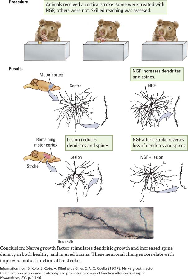
Behavioral therapy—
EXPERIMENT
Question: Does nerve growth factor stimulate recovery from stroke, influence neural structure, or both?

In principle, we might expect that any drug that stimulates the growth of new connections would help people recover from brain injury. However, that neural growth must occur in brain regions that can influence a lost function. A drug that stimulates synaptic growth on cells in the visual cortex, for example, would not enhance recovery of hand use. The visual neurons play no direct role in moving the hand.
A third strategy to generate new neural circuits uses either deep brain stimulation (DBS) or direct electrical stimulation of perilesional regions. The goal of electrical stimulation is to directly increase activity in remaining parts of specific damaged neural networks. In DBS, it is to put the brain into a more plastic (trainable) state so that rehabilitation therapies work better. Both strategies are in preliminary clinical trials.
Lost Neuron Replacement Solution
Focus 5-4 recounts a successful case of fetal stem cell transplantation. Section 13-2 describes SCN cell replacement.
The third strategy a patient like Donna could pursue is to generate new neurons to produce new neural circuits. The idea that brain tissue could be transplanted from one animal to another goes back a century. The evidence is good that tissue transplanted from fetal brains will grow and form some connections in the new brain.
The striatum, a region in the basal ganglia, includes the caudate nucleus and putamen.
Unfortunately, in contrast with transplanted hearts or livers, transplanted brain tissue functions poorly. The procedure seems most suited to conditions in which a small number of functional cells are required, as in the replacement of dopamine-
By 2004, dopamine-
Even in adults, neural stem cells line the subventricular zone, diagrammed in Figure 8-10A.
Adult stem cells offer a second way to replace lost neurons. Investigators know that the brain is capable of making neurons in adulthood. The challenge is to get the brain to do so after an injury. Brent Reynolds and Sam Weiss (1992) made the first breakthrough in this research.
Cells lining the ventricles of adult mice were removed and placed in a culture medium. The researchers demonstrated that if the correct trophic factors are added, the cells begin to divide and can produce new neurons and glia. Furthermore, if the trophic factors—
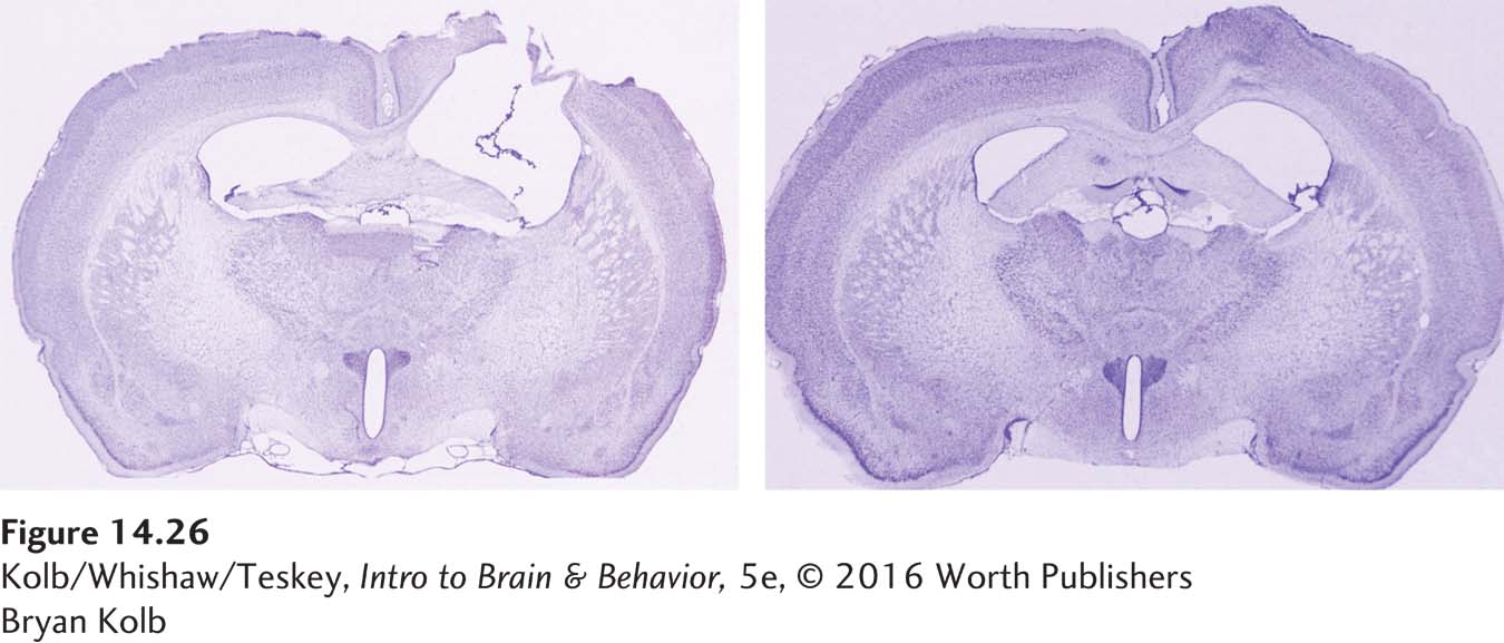
In principle, it ought to be possible to use trophic factors to stimulate the subventricular zone to generate new cells in the injured brain. If these new cells were to migrate to the site of injury and essentially regenerate the lost area, as shown in Figure 14-26, then it might be possible to restore at least some lost function. Not all lost behaviors could be restored, however, because the new neurons would have to establish the same connections with the rest of the brain that the lost neurons once had.

This task would be daunting: connections would have to be formed in an adult brain that already has billions of connections. Nonetheless, such a treatment might someday be feasible. Cocktails of trophic factors are effective in stimulating neurogenesis in the subventricular zone after brain injury. For example, in Figure 14-26, and many differentiated into immature neurons.
Although they did not integrate well into the existing brain, the new cells influenced behavior and led to functional improvement (Kolb et al., 2007). The mechanism of influence is poorly understood. The new neurons apparently had some trophic influence on the surrounding uninjured cortex. Preliminary clinical trials with humans are under way and so far show no ill effects in volunteers.
14-5 REVIEW
Recovery from Brain Injury
Before you continue, check your understanding.
Question 1
Three ways to compensate for the loss of neurons are (1) ____________, (2) ____________, and (3) ____________.
Question 2
Two ways of using electrical stimulation to enhance postinjury recovery are ____________ and ____________.
Question 3
Endogenous stem cells can be recruited to enhance functional improvement by using ____________ factors.
Question 4
What is the lesson of the three-
Answers appear in the Self Test section of the book.