6-5 Hormones
Berthold’s experiment demonstrated that hormones—chemicals released by endocrine glands into the bloodstream—
In 1849, European scientist A. A. Berthold removed a rooster’s testes and found that it no longer crowed, nor did it engage in sexual or aggressive behavior. Berthold then reimplanted one testis in the rooster’s body cavity. The rooster began crowing and displaying normal sexual and aggressive behavior again. The reimplanted testis did not establish any nerve connections, so Berthold concluded that it must release a chemical into the rooster’s circulatory system that influenced the animal’s behavior.
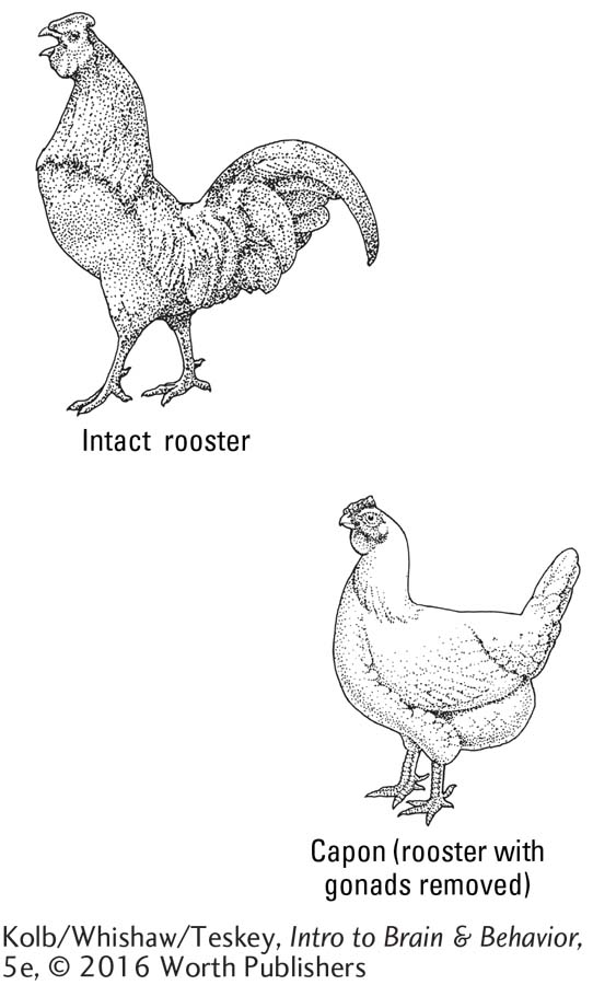
That chemical, we now know, is testosterone, the sex hormone secreted by the testes and responsible for the distinguishing characteristics of the male. The effect Berthold produced by reimplanting the rooster’s testis mimics the effect of administering testosterone to a castrated rooster, or capon. The hormone is sufficient to make the capon behave like a rooster.
We now know that many hormones affect the sexual characteristics and reproductive behavior of animals, including us humans. Hormonal effects are not limited to sexual behavior but also influence eating and drinking, growth, stress responses, and other bodily functions. Hormones are secreted by glands in the body and by the brain. Interacting brain and body hormones form feedback loops that regulate their activity. Hormonal influences change across the life-
Hierarchical Control of Hormones
Many hormones operate in a feedback system that includes the brain and the body. Figure 6-18 shows how the hypothalamus produces neurohormones that stimulate the pituitary gland to secrete releasing hormones into the circulatory system. The pituitary hormones in turn influence the remaining endocrine glands to release appropriate hormones into the bloodstream to act on various targets in the body and send feedback to the brain about the need for more or less hormone release.
Hormones not only affect body organs but also target virtually all aspects of brain function. Almost every neuron in the brain contains receptors on which various hormones can act. In addition to influencing sex organs and physical appearance, hormones affect neurotransmitter function, especially in neurons that influence sexual development and behavior (Barth et al., 2015). Hormones can influence gene expression by binding to special receptors on or in the cell, then being transported to the nucleus to influence gene transcription. Transcription in turn influences the synthesis of proteins needed for a variety of cellular processes. Thus, hormones influence brain and body structure and behavior.
Although many questions remain about how hormones produce or contribute to complex behavior, the diversity of their functions clarifies why the body uses hormones as messengers: their targets are so widespread that the best possible way of reaching all of them is to travel in the bloodstream, which goes everywhere in the body.
Classes and Functions of Hormones
Hormones can be used as drugs to treat or prevent disease. People take synthetic hormones as replacement therapy if the glands that produce the hormones are removed or malfunction. People also take hormones, especially sex hormones, to counteract the effects of aging, to increase physical strength and endurance, and to gain an advantage in sports. In the human body, as many as 100 hormones are classified chemically as either steroids or peptides.
Steroid hormones, such as testosterone and cortisol, are synthesized from cholesterol and are lipid (fat) soluble. Steroids diffuse away from their site of synthesis in glands, including the gonads, adrenal cortex, and thyroid. They bind to steroid receptors on the cell membrane or in the cell and frequently act on cellular DNA to influence gene transcription.
Peptide hormones, such as insulin, growth hormone, and the endorphins, are made by cellular DNA in the same way other proteins are made. They influence their target cell’s activity by binding to metabotropic receptors on the cell membrane, generating a second messenger that affects the cell’s physiology or gene transcription.
To refresh your understanding of metabotropic receptors, review Figure 5-15.
Steroid and peptide hormones fall into one of three main functional groups with respect to behavior, and they may function in more than one group:
Homeostatic hormones maintain a state of internal metabolic balance and regulate physiological systems. Mineralocorticoids (e.g., aldosterone) control both the concentration of water in blood and cells and the levels of sodium, potassium, and calcium in the body, and they promote digestive functions.
Page 202Gonadal (sex) hormones control reproductive functions. They instruct the body to develop as male (testosterone) or female (estrogen); influence sexual behavior and conception; and in women, control the menstrual cycle (estrogen and progesterone), birthing of babies, and release of breast milk (prolactin, oxytocin). These hormones, especially oxytocin, influence mother–
infant bonding, and in some species, including sheep, are essential for bonding to occur. Glucocorticoids (e.g., cortisol and corticosterone), a group of steroid hormones secreted in times of stress, are important in protein and carbohydrate metabolism and in controlling blood sugar levels and cellular absorption of sugar. Hormones activated in psychologically challenging events or emergencies prepare the body to cope by fighting or fleeing.
Homeostatic Hormones
Homeostasis comes from the Greek words stasis (standing) and homeo (in the same place).
Homeostatic hormones are essential to life. The body’s internal environment must remain within relatively constant parameters for us to function. An appropriate balance of sugars, proteins, carbohydrates, salts, and water is necessary in the blood, in the extracellular compartments of muscles, in the brain and other body structures, and in all cells. The internal environment must be maintained regardless of a person’s age, activities, or conscious state. As children or adults, at rest or in strenuous work, when we have overeaten or when we are hungry, to survive we need a relatively constant internal environment.
Normal glucose concentration in the bloodstream varies between 80 and 130 mg per 100 milliliters (about 3.3 oz) of blood.
A typical homeostatic function is controlling blood sugar level. After a meal, digestive processes result in increased glucose in the blood. One group of cells in the pancreas releases insulin, a homeostatic hormone that instructs the enzyme glycogen synthase in liver and muscle cells to start storing glucose in the form of glycogen. The resulting decrease in glucose decreases the stimulation of pancreatic cells so that they stop producing insulin, and glycogen storage stops. When the body needs glucose for energy, another hormone in the liver, glucagon, acts as a countersignal to insulin. Glucagon stimulates another enzyme, glycogen phosphorylase, to initiate glucose release from its glycogen storage site.
Diabetes mellitus is caused by a failure of the pancreatic cells to secrete enough insulin, or any at all. As a result, blood sugar levels can fall (hypoglycemia) or rise (hyperglycemia). In hyperglycemia, blood glucose levels rise because insulin does not instruct body cells to take up glucose. Consequently, cell function, including neuronal function, can fail through glucose starvation, even in the presence of high glucose levels in the blood. Chronic high blood glucose levels cause damage to the eyes, kidneys, nerves, heart, and blood vessels.
In hypoglycemia, inappropriate diet can lead to low blood sugar severe enough to cause fainting. Eric Steen and his coworkers (2005) propose that insulin resistance in brain cells may be related to Alzheimer disease. They raise the possibility that Alzheimer disease may be a third type of diabetes.
Hunger and eating are influenced by a number of homeostatic hormones, including leptin and ghrelin. Leptin (from the Greek for thin), secreted by adipose (animal fat) tissue, inhibits hunger and so is called the satiety hormone. Ghrelin (from the Indio-
Gonadal Hormones
We are prepared for our adult reproductive roles by the gonadal hormones that give us our sexual appearance, mold our identity on the continuum of male to female, and allow us to engage in sex-
The male Y chromosome contains a gene called the sex-
Section 8-4 explains how gonadal hormones participate in brain development.
The organizational hypothesis proposes that hormone action in the course of development alters tissue differentiation. Thus, testosterone masculinizes the brain early in life, having been taken up in brain cells, where it is converted into estrogen by the enzyme aromatase. Estrogen then acts on estrogen receptors to initiate a chain of events that includes activating certain genes in the cell nucleus. These genes contribute to the masculinization of brain cells and their interactions with other brain cells.
That estrogen, a hormone usually associated with the female, masculinizes the male brain may seem surprising. Estrogen does not have the same effect on the female brain, because females have a blood enzyme that binds to estrogen and prevents its entry into the brain. Hormones play a somewhat lesser role in producing the female body and brain, but they control the mental and physical aspects of menstrual cycles, regulate many facets of pregnancy and birth, and stimulate milk production for breastfeeding. In males, gonadal hormones demethylate, and so release, genes in the preoptic area of the hypothalamus to become active. The expression of these genes influences male sexual characteristics and behavior. Thus, active methylation of male sex–
Section 12-5 describes gonadal hormones’ effects on sexual behavior. Section 15-5 recounts sex differences in thinking patterns.
Gonadal hormones contribute to surprising differences in the brain and in cognitive behavior and play a role in male–
Three lines of evidence, summarized by Elizabeth Hampson and Doreen Kimura (2005), support the conclusion that sex-
Spatial and verbal tests given to females and males in many different settings and cultures show that males tend to excel in spatial tasks and females in verbal tasks.
Results of similar tests given to female participants in the course of the menstrual cycle show fluctuations in these test scores with phases of the cycle. During the phase in which the female sex hormones estradiol (metabolized from estrogen) and progesterone are at their lowest levels, women perform comparatively better on spatial tasks; during the phase in which levels of these hormones are high, women do comparatively better on verbal tasks.
Tests comparing premenopausal and postmenopausal women, women in various stages of pregnancy, and females and males with varying levels of circulating sex hormones all provide some evidence that hormones affect cognitive function.
Sex hormone–
Anabolic–Androgenic Steroids
A class of synthetic hormones related to testosterone has both muscle-
Synthetic steroid use rapidly spread to other countries and sports, eventually leading to a ban from track and field and then from many other sports, enforced by drug testing. Testing policy has led to a cat-
Today, the use of anabolic steroids is about equal among athletes and nonathletes. More than 1 million people in the United States have used anabolic steroids not only to enhance athletic performance but also to enhance physique and appearance. Anabolic steroid use in high schools may be as high as 7 percent for males and 3 percent for females.
The use of anabolic steroids carries health risks. Their administration results in the body reducing its manufacture of testosterone, which in turn reduces male fertility and spermatogenesis. Muscle bulk is increased and so is aggression. Cardiovascular effects include increased risk of heart attack and stroke. Liver and kidney function may be compromised, and the risk of tumors may increase. Male-
Anabolic steroids have approved clinical uses. Testosterone replacement is a treatment for hypogonadal males. It is also useful for treating muscle loss subsequent to trauma and for the recovery of muscle mass in malnourished people. In females, anabolic steroids are used to treat endometriosis and fibrocystic disease of the breast.
Glucocorticoids and Stress
Stress is a term borrowed from engineering to describe a process in which an agent exerts a force on an object. Applied to humans and other animals, a stressor is a stimulus that challenges the body’s homeostasis and triggers arousal. Stress responses, behavioral as well as physiological, include both arousal and attempts to reduce stress. A stress response can outlast a stress-
Activating a Stress Response
Surprisingly, the body’s response is the same whether the stressor is exciting, sad, or frightening. Robert Sapolsky (2004) uses the vivid image of a hungry lion chasing down a zebra to illustrate the stress response. The chase elicits divergent behavior in the two animals, but their physiological responses are identical. The stress response begins when the body is subjected to a stressor and especially when the brain perceives a stressor and responds with arousal, directed from the brain by the hypothalamus. The response consists of two separate sequences, one fast and the other slow.
THE FAST RESPONSE Shown at left in Figure 6-19, the sympathetic division of the ANS is activated to prepare the body and its organs for fight or flight. The parasympathetic division for rest and digest is turned off. The sympathetic division stimulates the medulla on the interior of the adrenal gland to release epinephrine. The epinephrine surge (often called the adrenaline surge after epinephrine’s original name) prepares the body for a sudden burst of activity. Among its many functions, epinephrine stimulates cell metabolism, readying the body’s cells for action.
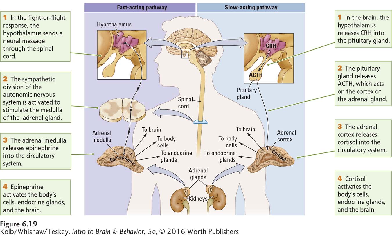
THE SLOW RESPONSE As shown at right in Figure 6-19, the slow response is controlled by the steroid cortisol, a glucocorticoid released from the outer layer (cortex) of the adrenal gland. Activating the cortisol pathway takes anywhere from minutes to hours. Cortisol has wide-
Ending a Stress Response
Normally, stress responses are brief. The body mobilizes its resources, deals with the challenge physiologically and behaviorally, and shuts down the stress response. Just as the brain is responsible for turning on the stress reaction, it is also responsible for turning it off. Consider what can happen if the stress response is not shut down:
The body continues to mobilize energy at the cost of energy storage.
Proteins are used up, resulting in muscle wasting and fatigue.
Growth hormone is inhibited, so the body cannot grow.
The gastrointestinal system remains shut down, reducing the intake and processing of nutrients to replace used resources.
Reproductive functions are inhibited.
The immune system is suppressed, contributing to the possibility of infection or disease.
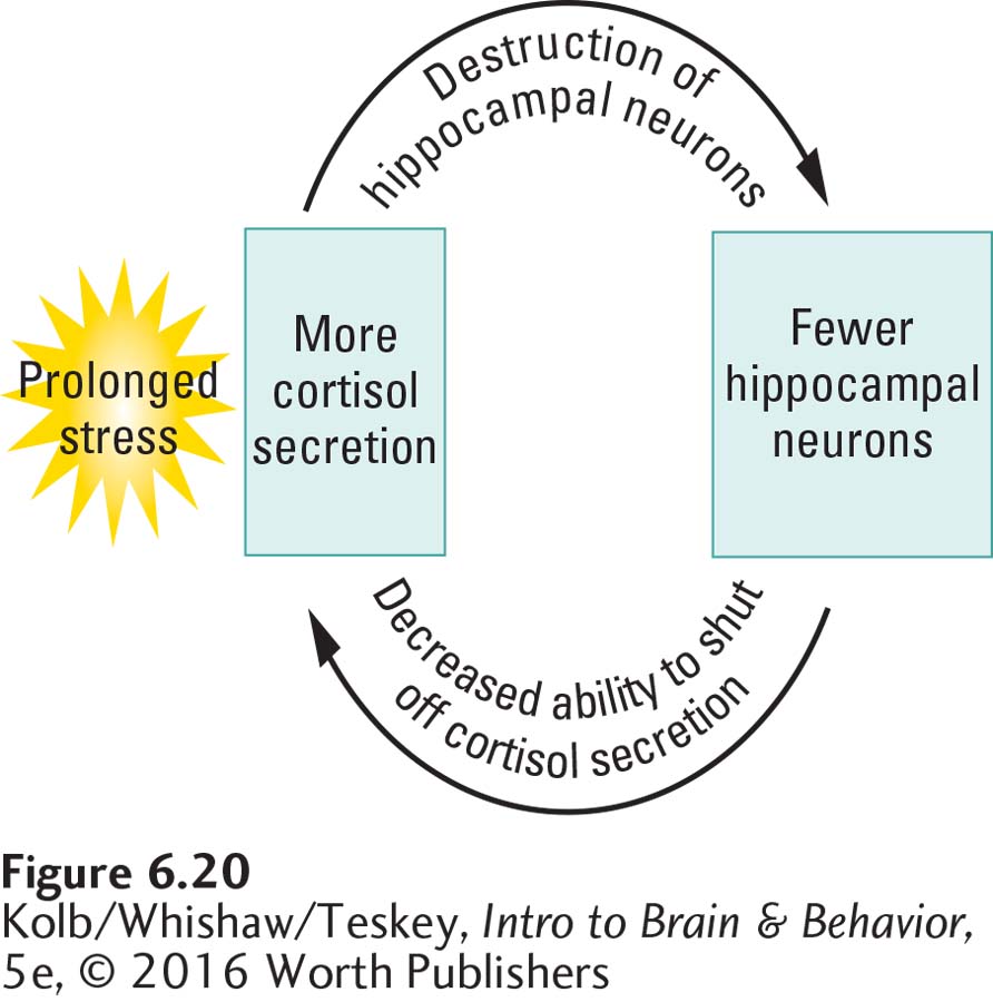
Sapolsky (2005) argues that the hippocampus plays an important role in turning off the stress response. The hippocampus contains a high density of cortisol receptors, and it has axons that project to the hypothalamus. Consequently, the hippocampus is well suited to detecting cortisol in the blood and instructing the hypothalamus to reduce blood cortisol levels.
There may, however, be a more insidious relation between the hippocampus and blood cortisol levels. Sapolsky observed wild-
Cortisol levels are usually regulated by the hippocampus, but if these levels remain elevated because a stress-
PTSD, introduced in Section 5-4 in relation to sensitization, is among the anxiety disorders detailed in Section 12-4. Focus 16-1 and Section 16-4 consider treatments.
Because stress response circuits in rats and monkeys are similar to those in humans, it is possible that excessive stress in humans can lead to similar brain changes. Because the hippocampus is thought to play a role in memory, stress-
Research has not yet determined whether the cumulative effects of stress damage the human hippocampus. For example, research on women who were sexually abused in childhood and were diagnosed with PTSD yields some reports of changes in memory or in hippocampal volume, as measured with brain imaging techniques. Other studies report no differences in abused and nonabused subjects (Landré et al., 2010). The fact that such apparently similar studies can obtain different results can be explained in several ways.
First, the amount of damage to the hippocampus that must occur to produce a stress syndrome is not certain. Second, brain imaging techniques may not be sensitive to subtle changes in hippocampal cell function or to moderate cell loss. Third, wide individual and environmental differences influence how people respond to stress. Fourth, neonatal stress can influence hippocampal neurogenesis (Lajud and Torner, 2015). The long-
Finally, humans are long-
The decrease in receptors and in glucocorticoid mRNA suggests that childhood abuse induces epigenetic changes in the expression of glucocorticoid genes. The decrease in glucocorticoid receptors presumably renders the hippocampus less able to depress stress responses. The importance of the McGowan study is its suggestion of a mechanism through which stress can influence hippocampal function without necessarily being associated with a decrease in hippocampal volume. This study further underscores the point that stress likely produces many changes in many brain regions. It is unlikely that all have been described or are understood (Clauss et al., 2015).
6-5 REVIEW
Hormones
Before you continue, check your understanding.
Question 1
The hypothalamus produces _______ that stimulate the _______ to secrete _______ into the circulatory system. Hormone levels circulating in the bloodstream send feedback to the _______.
Question 2
Hormones are classified chemically as _______ or _______.
Question 3
Broadly speaking, _______ hormones regulate metabolic balance; _______ hormones regulate reproduction; and _______ regulate stress.
Question 4
One class of synthetic hormones is _______, which increase _______ and have _______ effects.
Question 5
The stress response has a fast-
Question 6
Describe the proposed relationship among stress, cortisol, and the hippocampus.
Answers appear in the Self Test section of the book.