8-4 Brain Development and the Environment
Section 1-5 summarizes humanity’s acquisition of culture.
Developing behaviors are shaped not only by the maturation of brain structures but also by each person’s environment and experience. Neuroplasticity suggests that the brain is pliable and can be molded, at least at the microscopic level. Brains exposed to different environmental experiences are molded in different ways. Culture is an important aspect of the human environment, so culture must help to mold the human brain. We would therefore expect people raised in widely differing cultures to acquire brain structure differences that have lifelong effects on their behavior.
The brain is plastic in response not only to external events but also to events within a person’s body, including the effects of hormones, injury, and genetic mutations. The developing brain early in life is especially responsive to these internal factors, which in turn alter how the brain responds to external experiences. In this section, we explore a whole range of external and internal environmental influences on brain development. We start with a question: Exactly how does experience alter brain structure?
Experience and Cortical Organization
The Hebb synapse, diagrammed in Section 15-1, visualizes his predictions about synaptic plasticity. Section 14-4 elaborates Hebb’s contributions to learning theory.
Researchers can study the effects of experience on the brain and behavior by placing laboratory animals in different environments and observing the results. In one of the earliest such studies, Donald Hebb (1947) took a group of young laboratory rats home and let them grow up in his kitchen. A control group grew up in standard laboratory cages at McGill University.
The home-
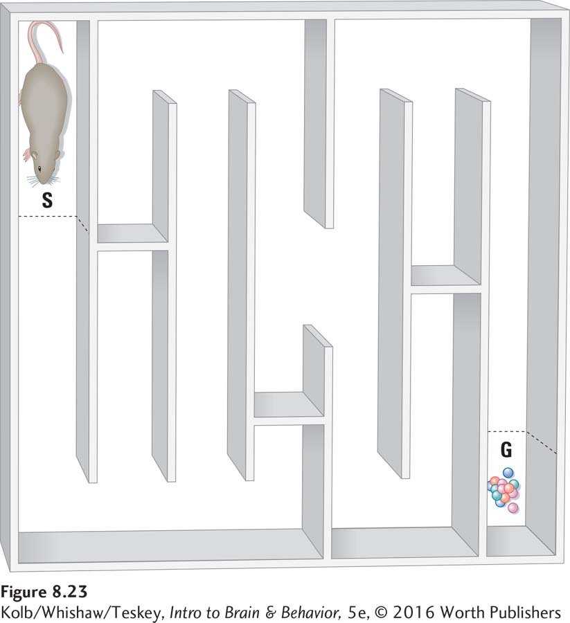
On the basis of his research, Hebb reasoned that people reared in a stimulating environment will maximize their intellectual development, whereas people raised in impoverished or under-
People living in slums typically have few formal educational resources—
Slum dwellers may not be well adapted for college life, however. This is probably closer to what Hebb had in mind when he referred to a slum environment as limiting intellectual potential. Indeed, Hebb’s logic led to the development of preschool television programs, such as Sesame Street, that offer enrichment for children who would otherwise have little preschool exposure to reading.
At 36 months of age, on average, the vocabulary of children from a low-
Seven decades ago, Hebb’s studies used complex stimulating environments, but much simpler experiences can also influence brain development. Tactile stimulation of human infants is important not only for bonding with caregivers but also for stimulating brain development. For example, tactile stimulation of premature infants in incubators speeds their growth and allows for quicker release from the hospital. Laboratory studies show that brushing infant rats for 15 minutes 3 times per day for the first 3 weeks of life also speeds up growth and development. The animals show enhanced motor and cognitive skills in adulthood as well. Tactile stimulation also dramatically improves recovery from brain injury incurred early in development.
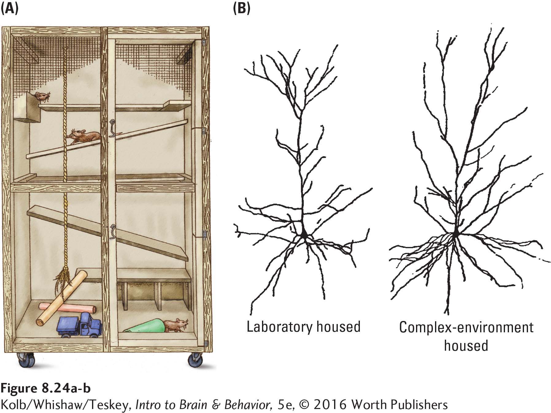
The idea that early experience can change later behavior seems sensible enough, but we are left to question why experience should make such a difference. One reason is that experience changes neuronal structure, which is especially evident in the cortex. Neurons in the brains of animals raised in complex environments, such as that shown in Figure 8-24A, are larger and richer in synapses than are those of animals reared in barren cages (Figure 8-24B). Similarly, 3 weeks of tactile stimulation increases synapse numbers all over the cortex in adulthood.
Focus 5-5 describes some structural changes neurons undergo as a result of learning.
Presumably, increased synapse numbers result from increased sensory processing in a complex and stimulating environment. The brains of animals raised in complex settings also display more (and larger) astrocytes. Although complex-
Figure 15-11 shows enhanced nerve tract connectivity in people with perfect pitch.
Like early exposure to language during development, early exposure to music alters the brain. Perfect (absolute) pitch, or the ability to re-
Such loss of plasticity does not mean that the adult human brain grows fixed and unchangeable. Adults’ brains are influenced by exposure to new environments and experiences, although more slowly and less extensively than children’s brains are. In fact, evidence reveals that experience affects the brain well into old age: good news for those of us who are no longer children.
Focus 6-2 details FASD.
It is becoming clear as well that prenatal events can modify brain development. The consensus is that perinatal adversity, such as gestational stress at or near birth, is a significant risk factor for later behavioral disorders (see Bock et al., 2014). Even events such as stress or drug use that occur before conception can lead to epigenetic effects in offspring. Examples include abnormalities in neural organization and behavior (e.g., Harker et al., 2015). Although such effects are usually presumed to come from maternal exposure before conception, increasing evidence from research on humans points to paternal preconception experience also modifying children’s brain development, perhaps including acquisition of fetal alcohol spectrum disorder (FASD).
8-3
Increased Cortical Activation for Second Languages
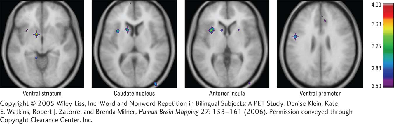
Most of the world’s population is bilingual, but people rarely learn their second language as early as their first. Denise Klein and her colleagues (2006) used both PET and fMRI to determine whether native and second languages differ in cortical activation.
The two languages overlap greatly in neural representation, but when participants are asked to repeat words, the second language shows greater activation in motor regions such as the striatum and cerebellum as well as in the frontal and temporal language regions. The investigators speculate that the second language places greater articulatory demands on the speaker. These demands correspond to the increased neural involvement in motor as well as language areas, as shown in the illustration.
Further support for Klein and colleagues’ conclusions from these imaging studies comes from a review of cortical mapping studies of bilingual patients undergoing neurosurgery. Carlo Giussani and his colleagues (2007) conclude that although all studies show language representation grossly located in the same cortical regions, distinct language-
Experience and Neural Connectivity
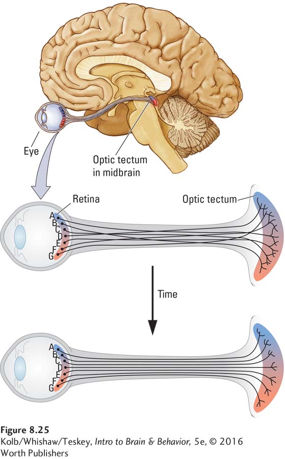
Experience can actually sculpt the brain prenatally, as studies of the developing visual system illustrate clearly. Consider the anatomical challenge of connecting the eyes to the rest of the visual system. A simple analogy will help. Imagine that students in a large lecture hall are each viewing the front of the room (the visual field) through a small cardboard tube, such as an empty paper towel roll. If each student looks directly ahead, he or she will see only a small bit of the total visual field.
Section 9-2 describes visual system anatomy. Figure 2-18 details midbrain structures.
Essentially, this is how the photoreceptor cells in the eyes act. Each cell sees only a small bit of the visual field. The problem is putting all of the bits together to form a complete picture. To do so, analogously to students sitting side by side, receptors that see adjacent views must send their information to adjacent regions in the various parts of the brain’s visual system, such as the midbrain. How do they accomplish this feat?
Roger Sperry (1963) suggested the chemoaffinity hypothesis, the idea that specific molecules in different cells in various midbrain regions give each cell a distinctive chemical identity. Each cell has an identifiable biochemical label. Presumably, incoming axons seek out a specific chemical, such as the tropic factors discussed in Section 8-2, and consequently land in the correct general midbrain region.
Many experiments have shown this process to take place prenatally as the eye and brain are developing. But the problem is that chemical affinity directs incoming axons only to a general location. To return to our two adjacent retinal cells, how do they now place themselves in the precisely correct position?
Here is where postnatal experience comes in: fine-
Now consider what happens to axons coming from different eyes. Although the neural inputs from the two eyes may be active simultaneously, cells in the same eye are more likely to be active together than are cells in different eyes. The net effect is that inputs from the two eyes tend to organize themselves into neural bands, called columns, that represent the same region of space in each eye, as shown on the left in Figure 8-26. Formation of these segregated cortical columns therefore depends on the patterns of coinciding electrical activity on the incoming axons.
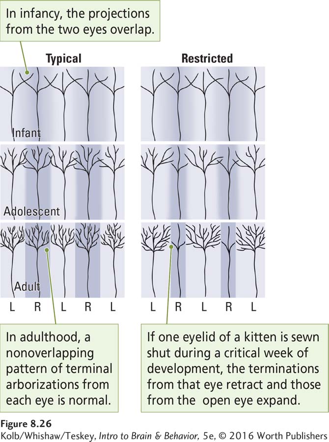
If experience is abnormal—
To summarize, an organism’s genetic blueprint is vague in regard to exactly which connections in the brain go to exactly which neurons. Experience fine-
Critical Periods for Experience and Brain Development
The preceding examples of perfect pitch and visual connectivity show that for healthy development, specific sensory experiences occurring at particular times are especially important. A time during which brain development is most sensitive to a specific experience is called either a critical period or a sensitive period.
The absence of appropriate sensory experience during a critical period may result in abnormal brain development, leading to abnormal behavior that endures even into adulthood. Our colleague Richard Tees offered an analogy to help explain the concept. He pictured the developing animal as a little train traveling past an environmental setting, perhaps the Rocky Mountains. All the windows are closed at the beginning of the journey (prenatal development), but at particular stages of the trip, the windows in certain cars open, exposing the occupants (different parts of the brain) to the outside world. Some windows open to expose the brain to specific sounds, others to certain smells, others to particular sights, and so on.
This exposure affects the brain’s development, and the absence of any exposure through an open window severely disturbs that development. As the journey continues, the windows become harder to open until finally they close permanently. This does not mean that the brain can no longer change, but changes become much harder to induce.
Now imagine two different trains, one headed through the Rocky Mountains and another, the Orient Express, traveling across Eastern Europe. The views from the windows are very different, and the effects on the brain are correspondingly different. In other words, not only is the brain altered by the experiences it has during a critical period, but the particular kinds of experiences encountered matter too.
An extensively studied, related behavior is imprinting, a critical period during which an animal learns to restrict its social preferences to a specific class of objects, usually the members of its own species. In birds, such as chickens and waterfowl, the critical period for imprinting often comes shortly after hatching. Typically, the first moving object a young hatchling sees is a parent or sibling, so the hatchling’s brain appropriately imprints to its own species.
Appropriate imprinting is not inevitable. Konrad Lorenz (1970) demonstrated that if the first animal or object that baby goslings encounter is a person, the goslings imprint to that person as though he or she were their mother. Figure 8-27 shows a flock of goslings that imprinted to Lorenz and followed him wherever he went. Incorrect imprinting has long-

Birds can imprint not just to humans but also to inanimate objects, especially moving objects. Chickens have been induced to imprint to a milk bottle sitting on the back of a toy train moving around a track. But the brain is not entirely clueless when it comes to selecting an imprinting target. Given a choice, young chicks will imprint on a real chicken over any other stimulus.
This quick acquisition and its permanent behavioral consequences suggest that during imprinting, the brain makes a rapid change of some kind, probably a structural change given the permanence of the new behavior. Gabriel Horn and his colleagues at Cambridge University (1985) tried to identify the changes in chicks’ brains during imprinting. The results of Horn’s electron microscopic studies show that synapses in a specific forebrain region enlarge with imprinting. Thus, imprinting seems a good model for studying brain plasticity during development, in part because the changes are rapid, related to specific experience, and localized in the brain.
As noted in Section 8-3, brain development can be affected by either parent’s experiences before conception or those of the mother or fetus during gestation. Because developmental events change so dramatically and quickly in utero, we should not be surprised that the effects of fetal experiences vary with the precise developmental stage. As a rule, the CNS is especially sensitive during gestational weeks 4 to 8, as the neural tube forms. It remains sensitive through the period of cerebral neurogenesis, which continues until the end of the second trimester. Robbin Gibb and her colleagues (2014) have shown that housing pregnant rats in complex environments, as in Figure 8-24A, results in the offspring showing increased dendritic spine density in the cortex, as though the animals had been placed in the environment in adulthood.
Abnormal Experience and Brain Development
If complex or enriched experiences can stimulate brain growth and influence later behavior, severely restricted experiences seem likely to retard both brain growth and behavior. To study the effects of such restrictions, Donald Hebb and his colleagues (Clarke et al., 1951) placed young Scottish terriers in the dark with as little stimulation as possible and compared their behavior to that of dogs raised in a typical environment.
When the dogs raised in the barren environment, obviously unethical by today’s standards, were later removed from it, their behavior was highly unusual. They showed virtually no reaction to people or other dogs and appeared to have lost any pain sensation. Even sticking pins in them (equally unethical) produced no response. When given a dog version of the Hebb–
Results of subsequent studies show specifically that depriving young animals of visual input or of maternal contact has devastating consequences for their behavioral development and presumably for their brain development. Austin Riesen (1982) and his colleagues extensively studied animals raised in the dark. They found that even though the animals’ eyes still work, they may be functionally blind after early visual deprivation. An absence of visual stimulation results in the atrophy of dendrites on cortical neurons, essentially the opposite of the results observed in the brains of animals raised in complex and stimulating environments.
Not only does the absence of specific sensory inputs adversely affect brain development; so do more complex atypical experiences. In the 1950s, Harry Harlow (1971) began the first systematic laboratory studies of analogous deprivation in laboratory animals. Harlow showed that infant monkeys raised without maternal (or paternal) contact develop grossly atypical intellectual and social behaviors in adulthood.
Harlow separated baby monkeys from their mothers shortly after birth and raised them in individual cages. Perhaps the most stunning effect occurred in adulthood, when these animals were totally unable to establish normal relations with other animals. Unfortunately, Harlow did not analyze the deprived monkeys’ brains. We would predict atrophy of cortical neurons, especially in the frontal lobe regions related to social behavior. Harlow’s student Stephen Suomi continues to study early experiences in monkeys at the U.S. National Institute of Child Health and Human Development. He has found a wide variety of hormonal and neurological abnormalities among motherless monkeys, including epigenetic changes (see the review by Suomi, 2011).
Children in a barren environment or abused or neglected are at a serious disadvantage later in life. Proof is the hampered intellectual and motor development displayed by children raised in dreadful circumstances such as those described in Clinical Focus 8-4, Romanian Orphans, on page 272. Although some argue that children can succeed in school and in life if they really want to, abnormal developmental experiences can clearly alter the brain irrevocably. As a society, we cannot be complacent about the environment to which our children are exposed.
Early exposure to stress, including prenatally, also has major effects on a child’s later behavior. Stress can alter the expression of certain genes, such as those related to serotonin (5-
Section 6-5 explains the neurobiology of the stress response. Section 16-4 connects mood and reactivity to stress.
Stress early in life may predispose people to develop behavioral disorders, such as depression (Sodhi & Sanders-
Prenatal experiences also can lead to abnormal behavior in children and adults. Exposing the fetus to alcohol, especially in the first two trimesters, can lead to FASD, as can excessive alcohol use by either parent before conception. Similarly, children exposed in utero to radiation in the aftermath of the 1986 accident at the Chernobyl nuclear power plant later developed a range of cognitive disorders, including lowered IQ scores. Effects were most severe if they had been exposed during gestational weeks 8 to 25, the critical period of cerebral neurogenesis (Nyagu et al., 1998).
Hormones and Brain Development
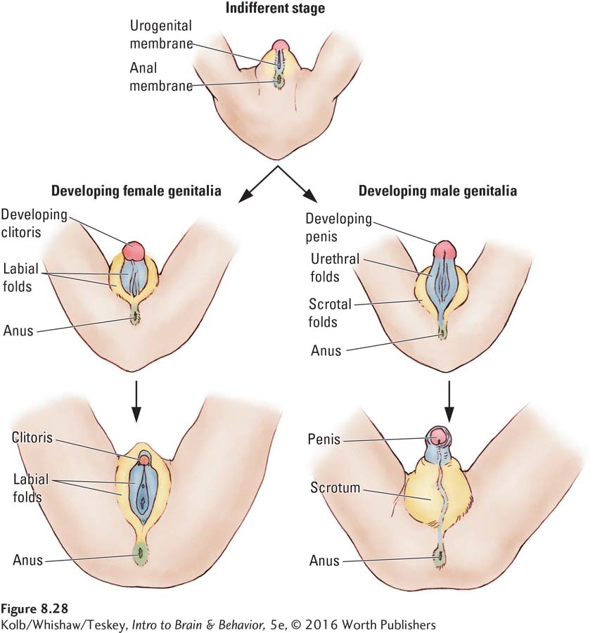
The determination of sex is largely genetic. In mammals, the Y chromosome in males controls the process by which an undifferentiated, primitive gonad develops into testes, illustrated in Figure 8-28. The genitals begin to form in the seventh week after conception, but they appear identical (indifferent) in the two sexes at this early stage. No sexual dimorphism, or structural difference, yet exists. The testes subsequently secrete the sex hormone testosterone, which stimulates development of male reproductive organs and later, in puberty, the appearance of male secondary sexual characteristics such as facial hair and deepening of the voice.
8-4
Romanian Orphans
In the 1970s, Romania’s Communist regime outlawed all forms of birth control and abortion. The natural result was more than 100,000 unwanted children in state-
The children were housed and clothed but given virtually no environmental stimulation. Mostly they were confined to cots with few, if any, playthings and virtually no personal interaction with overworked caregivers, who looked after 20 to 25 children at once. Bathing often consisted of being hosed down with cold water.
After the Communist government fell, the outside world intervened. Hundreds of these children were placed in adoptive homes throughout the world, especially in the United States, Canada, and the United Kingdom. Studies of these severely deprived children on arrival in their new homes document malnourishment, chronic respiratory and intestinal infections, and severe developmental impairments.
A British study by Michael Rutter (1998) and his colleagues assessed the orphans at two standard deviations below age-
The improvement these children showed in the first 2 years after placement in their adoptive homes was nothing short of spectacular. Average height and weight advanced to nearly normal, although head circumference remained below normal. Many tested in the normal range of motor and cognitive development. But a significant number were still considered intellectually impaired. What caused these individual differences in recovery from the past deprivation?
The key factor was age at adoption. Children adopted before 6 months of age did significantly better than those adopted later. In a Canadian study by Elenor Ames (1997), Romanian orphans who were adopted before 4 months of age and then tested at age 4½ had an average Stanford–
Charles Nelson and his colleagues (Berens & Nelson, 2015; Nelson et al., 2007; Smyke et al., 2012) analyzed cognitive and social development as well as event-
The inescapable conclusion is that the human brain may be able to recover from a brief period of extreme deprivation in early infancy, but periods longer than 24 months produce significant developmental abnormalities that cannot be overcome completely. The studies of Romanian orphans make clear that the developing brain requires stimulation for healthy development. Although the brain may be able to catch up after a brief deprivation, severe deprivation lasting many months results in a small brain and associated behavioral abnormalities, especially in cognitive and social skills.

Gonadal (sex) hormones change the genetic activity of certain cells, most obviously those that form the genitals, but neural cells also respond to them. Regions of the embryonic brain thus also may begin to show sexual dimorphism as testosterone secretion begins, about 60 days after conception. What does sexual differentiation have to do with brain development? Although the answer is largely hormonal, genetic influences contribute, too.
Sections 6-5 and 12-5 detail the actions of gonadal hormones, including testosterone.
Testosterone stimulates sexual differentiation in male embryos. In its absence, female embryos develop. Prenatal exposure to gonadal hormones shapes male and female brains differently, because these hormones activate different genes in the two sexes. Experience, then, affects male and female brains differently. Clearly, genes and experience begin to shape the developing brain very early.
Gonadal Hormones and Brain Development
Testosterone, the best-
Testosterone does not affect all body organs or all brain regions, but it does affect many brain regions in many ways. It affects the number of neurons formed in certain brain areas, reduces the number of neurons that die, increases cell growth, increases or reduces dendritic branching and synaptic growth, and regulates synaptic activity, among other effects.
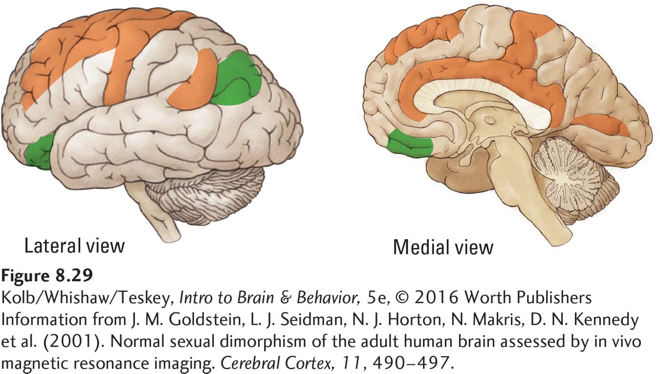
Estrogens, the sex hormones responsible for the female’s distinguishing characteristics, also probably influence postnatal brain development. Jill Goldstein and her colleagues found sex differences in the volume of cortical regions known to have differential levels of receptors for testosterone (androgen receptors) and estrogen, respectively, as shown in Figure 8-29 (Goldstein et al., 2001). Orange areas in the figure are larger in females, and green areas are larger in males. Clearly, a male brain and a female brain are not the same. Hormones alter brain development, and clear sex differences appear in the rate of brain development (see Figure 8-19).
Testosterone’s effects on brain development were once believed unimportant, because this hormone was thought primarily to influence brain regions related to sexual behavior, not regions of higher functions. This belief is false. Testosterone changes cell structure in many cortical regions, with diverse behavioral consequences that include influences on cognitive processes.
Jocelyne Bachevalier adapted her method, shown in Experiment 8-1 on page 264, by training infant male and female monkeys in the concurrent discrimination task. The animal has to learn which of two objects in a series of pairs conceals a food reward. Bachevalier also trained the animals in another task, object reversal learning. The task is to learn that one object always conceals a food reward, whereas another object never does. After the animal learns this pattern, the reward contingencies are reversed so that the particular object that has always been rewarded is now never rewarded, and the formerly unrewarded object now conceals the reward. When the animal learns this new pattern, the contingencies are reversed again, and so on for five reversals.
Bachevalier found that 2½-month-
Bachevalier and her colleague William Overman (Overman et al., 1996) repeated the experiment with children 15 to 30 months old. The results were the same: boys were superior at the object reversal task and girls were superior at the concurrent task. The investigators found no such male–
Lifelong Effects of Gonadal Hormones
Although gonadal hormones’ biggest effects on the brain may come during early development, their role there is by no means finished in infancy. Both testosterone and estrogen (which females’ ovaries produce in large quantities) continue to influence brain structure throughout an animal’s life. In fact, removal of the ovaries in middle-
Gonadal hormones also affect how the brain responds to environmental events. For instance, among rats housed in complex environments, males show more dendritic growth in neurons of the visual cortex than do females (Juraska, 1990). In contrast, females housed in this setting show more dendritic growth in the hippocampus than males do. Apparently, the same experience can affect the male and female brain differently owing to the mediating influence of gonadal hormones.
As females and males develop, then, their brains continue to diverge more and more, much like a fork in a road. After you set out on one path, your direction is forever changed, as the roads increasingly course farther apart.
Details on sexual orientation and gender identity appear in Section 12-5.
To summarize, gonadal hormones alter basic neuronal development, shape experience-
In part, it is true that environmental factors exert a major influence. But one reason they do so may be that male and female brains are different to start with. Even the same events experienced by structurally different brains may lead to different effects on those brains. Evidence shows that significant experiences, such as prenatal stress, produce markedly different changes in gene expression in the frontal cortex of male and female rats (Mychasiuk et al., 2011).
Another key question related to hormonal influences on brain development is whether any sex differences in brain organization might be independent of hormonal action. In other words, are differences in the action of sex chromosome genes unrelated to sex hormones? Although little is known about such genetic effects in humans, studies of birds clearly show that genetic effects on brain cells may indeed contribute to sex differentiation.
Songbirds have an especially interesting brain dimorphism: in most species, males sing and females do not. This behavioral difference between the sexes is directly related to a neural birdsong circuit present in males but not in females. Robert Agate and his colleagues (2003) studied the brain of a rare gynandromorph zebra finch, shown in Figure 8-30. This bird exhibits physical characteristics of both sexes.
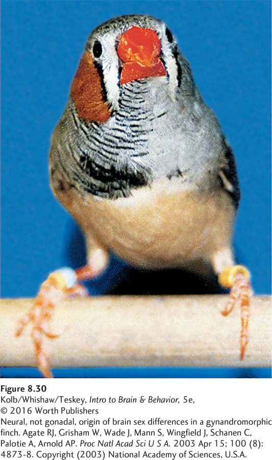
Genetic analysis shows that cells on one half of the bird’s brain and body are genetically female and on the other half are genetically male. The two sides of the gynandromorph’s body and brain were exposed to the same hormones during prenatal development. Thus, the effect of male and female genes on the birdsong circuit can be examined to determine how the genes and hormones might interact.
If the sex difference in the birdsong circuit were totally related to the presence of hormones prenatally, then the two sides of the brain should be equally masculine or feminine. Agate’s results confirm the opposite: the neural song circuit is masculine on the male side of the brain. Only a genetic difference that was at least partly independent of hormonal effects could explain such a structural difference in the brain.
Adolescent Onset of Mental Disorders
Adolescence is a time of rapid brain change related both to pubertal hormones and to psychosocial stress. Relationships with parents and peers are among the prime stressors, as is school. Add to this the finding, charted in Figure 8-31, that the peak age of onset for any mental disorder is estimated at 14 years (Paus et al., 2008).
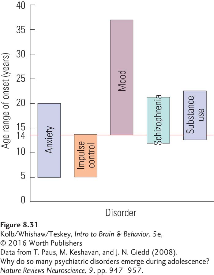
Figure 8-31 reveals that age of onset differs across disorders. However, anxiety disorders, psychoses (including schizophrenia), bipolar disorder, depression, eating disorders, and substance abuse most commonly emerge by or during adolescence. From an evolutionary perspective, neurobiological and associated behavioral changes linked with the period we define as adolescence are designed to optimize the brain for challenges that lie ahead in adulthood. But the brain’s plasticity in adolescence can also make it vulnerable to psychopathologies that can endure for the rest of the individual’s life.
Gut Bacteria and Brain Development
We have emphasized factors that affect CNS development directly, but a less direct route exerts itself via the enteric nervous system. The ENS sends information to the brain that affects our mental state. The brain in turn can modify gut function.
An important component of the ENS is the microbiome, the bacteria in the gut with which the ENS interacts. About 1014 microbiota populate the adult gut, which means that microbiota outnumber the host body cells by a factor of 10. But in utero, the fetus’s gut is sterile. It is only at birth that trillions of microbes from the mother’s vaginal and anal fluids, and later from her skin, invade the baby’s body and start to grow.
Section 2-5 introduces the ENS and microbiome.
Many neurodevelopmental disorders, including autism, may be related to an atypical microbiome early in life (e.g., Finegold et al., 2012). Elaine Hsiao and her colleagues (2013) studied a mouse model known to display features of ASD. These mice produce few social auditory vocalizations, about one-
Injury and Brain Development
Dating to the late 1800s, infants and children were generally believed to show better recovery from brain injury than adults. In the 1930s, Donald Hebb studied children with major birth-
Have other studies confirmed Hebb’s conclusion? Few anatomical studies of humans with early brain injuries exist, but we can make some general predictions from studying laboratory animals. In general, early brain injuries do produce atypical brains, especially at certain critical periods in development.
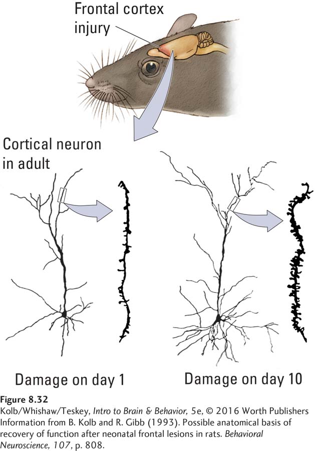
For humans, the worst time appears to be in the last half of the intrauterine period and the first couple of months after birth. Rats and cats that are injured at a comparable time have a significantly smaller brain than average, and their cortical neurons show general atrophy relative to healthy brains, as illustrated on the left in Figure 8-32. Behaviorally, these animals appear cognitively deficient over a wide range of skills.
Injury to the developing brain is not always devastating. For example, researchers have known for more than 100 years that children with brain injuries in the first couple of years after birth almost never have the severe language disturbances common to adults with equivalent injuries. Animal studies help explain why.
Whereas damage to the rat brain in the developmental period comparable to the last few months of gestation in humans produces widespread cortical atrophy, damage at a time in rat brain development roughly comparable to ages 6 months to 2 years in humans actually produces more dendritic development in rats (Figure 8-32 above). Furthermore, these animals show dramatic recovery of functions, which implies that during development the brain has a capacity to compensate for injury. Parallel studies in cats have shown extensive reorganization of cortex-
Drugs and Brain Development
The U.S. National Institute on Drug Abuse (2012) estimates that 16 percent of babies born alive in the United States today are exposed to nicotine in utero. Similar statistics on alcohol consumption by pregnant mothers are not available, but the effects of alcohol on the fetus are well documented. Even low doses of commonly prescribed drugs, including antidepressants, antipsychotics, and pain-
Section 6-3 reviews nicotine’s prominence as a gateway drug.
NIDA also estimates that 5.5 percent of expectant mothers, approximately 221,000 pregnant women each year in the United States, use an illicit drug at least once in the course of their pregnancy. Among pregnant teenagers aged 15 to 17, that figure climbs to 16%, or about 14,000 women. And what about caffeine and nicotine? More than likely most children were exposed to caffeine (from coffee, tea, cola, energy drinks, and chocolate) in utero and about 16% were exposed to nicotine. Laboratory animal studies have shown that prenatal exposure to nicotine alters the brain’s response to complex housing: the brain appears less plastic (e.g., Mychasiuk et al., 2014).
Focus 7-4 details ADHD and Focus 14-1 details dyslexia.
The precise effects of prenatal drug intake on brain development are poorly understood, but the overall conclusion from current knowledge is that children with prenatal exposure to a variety of psychoactive drugs have an increased likelihood of later drug use (e.g., Minnes et al., 2014). Although, again, childhood disorders are poorly studied, many experts suggest that they—
Other Sources of Abnormal Brain Development
The nervous system need not be damaged by external forces to develop abnormally. Many genetic aberrations are believed to result in abnormalities in brain development and ultimately brain structure. Spina bifida, in which the genetic blueprint goes awry and the neural tube does not close completely, leads to an incompletely formed spinal cord. After birth, unless treated with folic acid, children with spina bifida usually have serious motor problems.
Imagine what happens if some genetic aberration causes improper closure of the front end of the neural tube. Because the front end of the neural tube forms the brain (see Figure 8-5), this failure results in gross abnormalities in brain development known as anencephaly. Affected infants die soon after birth.
Atypical brain development can be much subtler than anencephaly. For example, if cells do not migrate to their correct locations, and if these mispositioned cells do not subsequently die, they can disrupt brain function and may lead to disorders ranging from seizures to schizophrenia (see review by Guerrini et al., 2007). In a variety of conditions, neurons fail to differentiate normally. In certain cases, neurons fail to produce long dendrites or spines, which results in abnormal brain connectivity and developmental disabilities.
The opposite condition also is possible: neurons continue to make dendrites and form connections with other cells until the neurons are extraordinarily large. The functional consequences of all the newly formed connections can be devastating. Excitatory synapses in the wrong location effectively short-
Subtle abnormal events also can be devastating, even terminal. Sudden infant death syndrome (SIDS), the unexplained death while asleep of a seemingly healthy infant less than 1 year old, kills about 2500 babies yearly in the United States alone. Postmortem studies reveal that SIDS victims are more likely than other babies to have a particular gene variation that makes the serotonin transporter unusually efficient. Normally, the serotoninergic system helps to stimulate a respiratory mechanism that responds to high carbon dioxide levels in the blood and acts to expel the gas.
In babies who die of SIDS, serotonin is cleared from the synapse more rapidly than normal. This action makes 5-
In addition to the serotonin transporter abnormality, David Paterson and his colleagues (2006) found an abnormally low occurrence of 5-
A curious consequence of abnormal brain development is that behavioral effects may emerge only as the brain matures and the maturing regions begin to play a greater role in behavior. This consequence is true especially of frontal lobe injuries. The frontal lobes continue to develop into early adulthood (see Figure 8-17), and often not until adolescence do the effects of frontal lobe abnormalities become noticeable.
Section 16-4 describes the schizophrenic brain and Section 5-3 a possible relation to excessive DA or 5-
Schizophrenia is a disease characterized by its slow development, usually not becoming obvious until late adolescence. Clinical Focus 8-5, Schizophrenia on page 278, relates disease progress and its possible origin.
8-5
Schizophrenia
When Mrs. T. was 16 years old, she began to experience her first symptom of schizophrenia: a profound feeling that people were staring at her. These bouts of self-
At first Mrs. T.’s illness was intermittent, and the return of her intelligence, warmth, and ambition between episodes allowed her to complete several years of college, to marry, and to rear three children. She had to enter a hospital for her illness for the first time at age 28, after the birth of her third child, when she began to hallucinate.
Now, at 45, Mrs. T. is never entirely well. She has seen dinosaurs on the street and live animals in her refrigerator. While hallucinating, she speaks and writes in an incoherent, but almost poetic way. At other times, she is more lucid, but even then the voices she hears sometimes lead her to do dangerous things, such as driving very fast down the highway in the middle of the night, dressed only in a nightgown. . . . At other times and without any apparent stimulus, Mrs. T. has bizarre visual hallucinations. For example, she saw cherubs in the grocery store. These experiences leave her preoccupied, confused, and frightened, unable to perform such everyday tasks as cooking or playing the piano. (Gershon & Rieder, 1992, p. 127)
It has always been easier to identify schizophrenic behavior than to define schizophrenia. Perhaps the one universally accepted criterion for its diagnosis is the absence of other neurological disturbances or affective (mood) disorders that could cause a person to lose touch with reality—
Symptoms of schizophrenia vary, suggesting that biological abnormalities also vary from person to person. Most patients appear to stay at a fairly stable level after the first few years of symptoms, with little evidence of a decline in neuropsychological functioning. Symptoms come and go, much as for Mrs. T., but the severity is relatively constant after the first few episodes.
Numerous studies have investigated the brains of schizophrenia patients, both in MRI and CT scans and in autopsies. Although the results vary, most neuroscientists agree that schizophrenic brains weigh less than normal and have enlarged ventricles. Research findings also suggest that brains affected by schizophrenia have smaller frontal lobes (or at least a reduction in the number of neurons in the prefrontal cortex) and thinner parahippocampal gyri.
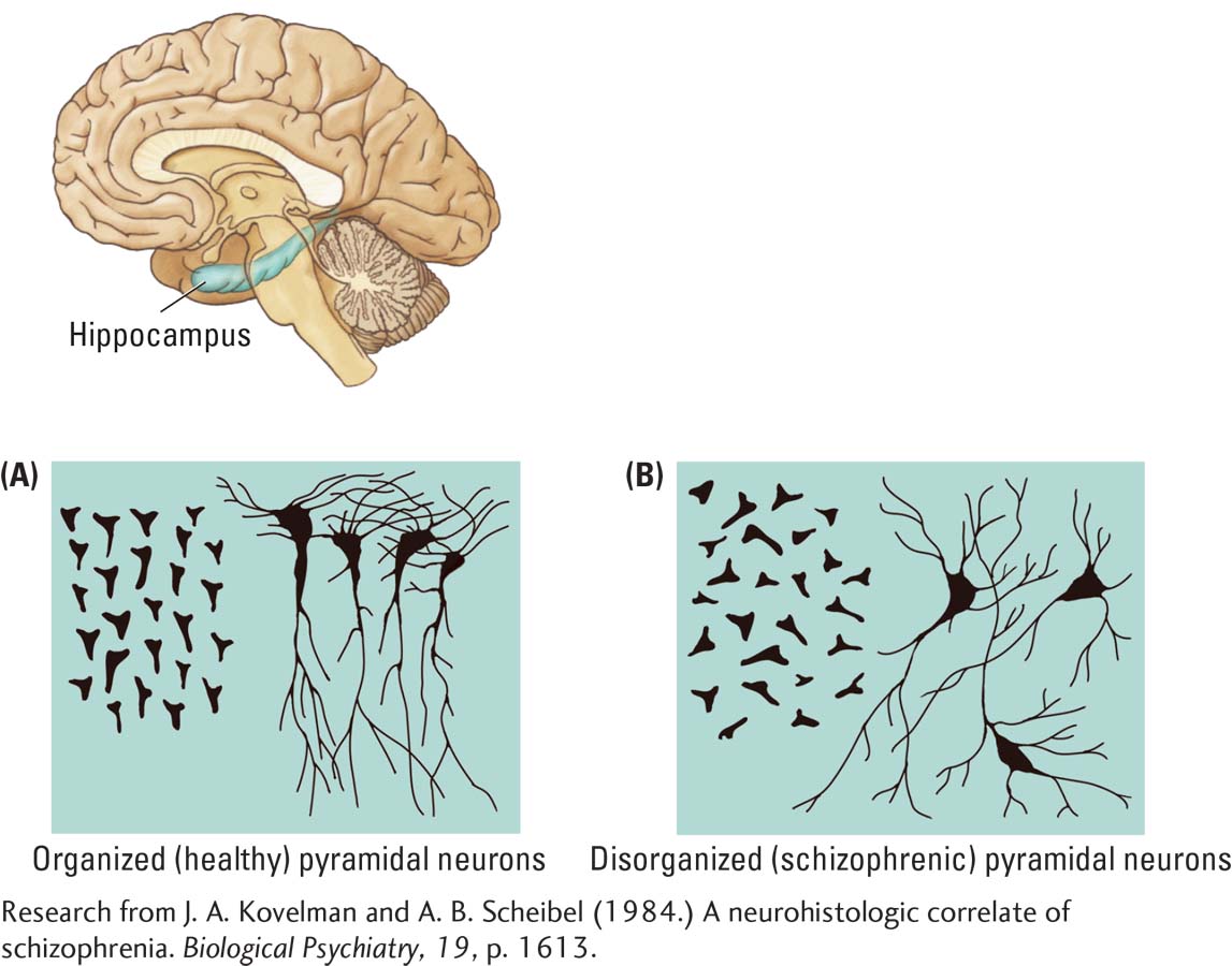
Joyce Kovelman and Arnold Scheibel (1984) found abnormalities in the orientation of hippocampal neurons in people with schizophrenia. Rather than the consistently parallel orientation of neurons in this region characteristic of healthy brains, schizophrenic brains have a more haphazard organization, as shown in the accompanying drawings.
Evidence is increasing that the abnormalities observed in schizophrenic brains are associated with disturbances of brain development. William Bunney and his colleagues (1997) suggested that at least a subgroup of those with schizophrenia underwent either environmental insults or some type of abnormal gene activity in the fourth to sixth month of fetal development.
These events are thought to result in abnormal cortical development, particularly in the frontal lobes. Later in adolescence, as the frontal lobes approach maturity, the person begins to have symptoms deriving from this abnormal prenatal development.
Developmental Disability
Figure 3-22 illustrates trisomy, the chromosomal abnormality that causes Down syndrome.
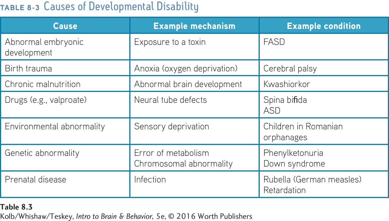
Impaired cognitive functioning accompanies abnormal brain development. Impairment may range from mild, allowing an almost normal lifestyle, to severe, requiring constant care. As summarized in Table 8-3, such developmental disability can result from chronic malnutrition, genetic abnormalities such as Down syndrome, hormonal abnormalities, brain injury, or neurological disease. Different causes produce different abnormalities in brain organization, but the critical similarity across all types of developmental disability is that the brain is not normal.
Dominique Purpura (1974) conducted one of the few systematic investigations of developmentally disabled children’s brains. Purpura used Golgi stain to examine the neurons of children who had died of accident or disease unrelated to the nervous system. When he examined the brains of children with various forms of intellectual disability, he found that dendrite growth was stunted and the spines very sparse relative to dendrites from children of typical intelligence, as illustrated in Figure 8-33.
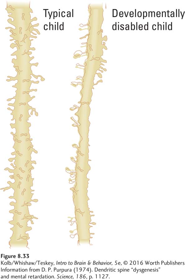
The simpler neuronal structure probably indicates a marked reduction in the number of brain connections, which presumably caused the developmental disability. Variation in both the nature and the extent of neuronal abnormality in different children would lead to different behavioral syndromes.
8-4 REVIEW
Brain Development and the Environment
Before you continue, check your understanding.
Question 1
The idea that specific molecules in different cells in various midbrain regions give each cell a distinctive chemical identity is known as the __________.
Question 2
Subnormal visual stimulation to one eye during early development can lead to a loss of acuity, known as __________.
Question 3
The hormone __________ masculinizes the brain during development.
Question 4
The brain’s sensitivity to experience is highest during __________.
Question 5
Why do so many mental disorders appear during adolescence?
Answers appear in the Self Test section of the book.