Chapter Introduction
How Do We Study the Brain’s Structures and Functions?
RESEARCH FOCUS 7-
LINKING NEUROANATOMY AND BEHAVIOR
EXPERIMENT 7-
METHODS OF BEHAVIORAL NEUROSCIENCE
MANIPULATING BRAIN–
RECORDING ACTION POTENTIALS FROM SINGLE CELLS
EEG: RECORDING GRADED POTENTIALS FROM THOUSANDS OF CELLS
MAPPING BRAIN FUNCTION WITH EVENT-
CLINICAL FOCUS 7-
MAGNETOENCEPHALOGRAPHY
FUNCTIONAL MAGNETIC RESONANCE IMAGING
OPTICAL TOMOGRAPHY
POSITRON EMISSION TOMOGRAPHY
MEASURING BRAIN CHEMISTRY
MEASURING GENES IN BRAIN AND BEHAVIOR
CLINICAL FOCUS 7-
EPIGENETICS: MEASURING GENE EXPRESSION
BENEFITS OF ANIMAL MODELS OF DISEASE
ANIMAL WELFARE AND SCIENTIFIC EXPERIMENTATION
RESEARCH FOCUS 7-

7-1
Tuning In to Language
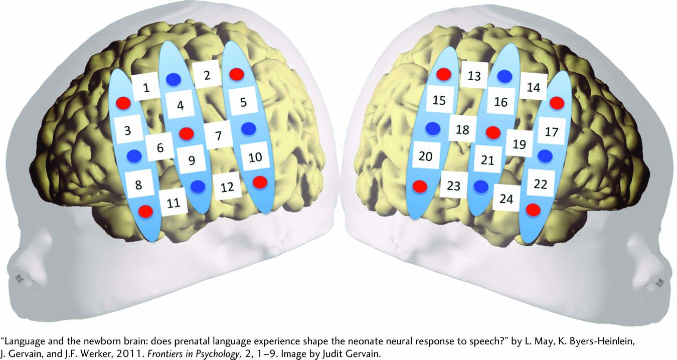
The continuing search to understand the organization and operation of the human brain is driven largely by emerging technologies. Over the past decade, neuroscience researchers have developed dramatic new noninvasive ways to image the brain’s activity in people who are awake. One technique, functional near-
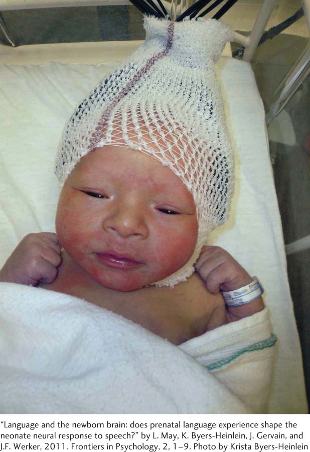
fNIRS allows investigators to measure oxygen consumption as a surrogate marker of neuronal activity in relatively select cortical regions, even in newborn infants. In one study (May et al., 2011), newborns (0–
When newborns listened to a familiar language, their brain showed a general increase in oxygenated hemoglobin; when they heard an unfamiliar language, oxygenated hemoglobin decreased overall. But when the babies heard the same sentences played backward, there was no difference in brain response to either language.
The opposing responses to familiar and unfamiliar languages mean that prenatal language experience shapes how the newborn brain responds to familiar and unfamiliar tongues. This finding leads to many questions. Among them: How does prenatal exposure to language influence later language learning? Do children who are exposed to multiple languages prenatally show better language acquisition than those exposed to just one? How much prenatal language exposure is necessary, and do premature infants show the same results as full-
Whatever the answers, this study shows that the prenatal brain is tuned in to the language environment into which it will be born.
The simple, noninvasive nature of fNIRS likely will yield new insights not only into brain development but also into adult brain function. Over the coming decades, our understanding of the brain–
To understand how far neuroscience research methods have progressed, imagine that it is the year 1800. You are a neurologist interested in studying how the brain works. The challenge is how to begin. The two most obvious choices are to dissect the brains of dead people and other animals or to study people who have sustained a brain injury. Indeed, this was how the relationship between brain and behavior was studied well into the twentieth century.
Section 4-1 reviews how the EEG enabled investigators to explain electrical activity in the nervous system.
Techniques for studying the brain’s physiological processes emerged in the years between World War I and World War II. One breakthrough was the electroencephalograph (EEG), developed for humans by Hans Berger in the 1930s. Advances in understanding genetics and the analysis of behavior in the early 1950s set the stage for phenomenal advances in neuroscience knowledge. Scientists recognize that new technologies allow for novel insights, lead to more questions, and can dramatically advance their discipline. A large number of Nobel Prizes have been awarded for the development and implementation of new technologies.
Today, brain–
We begin this chapter by reviewing how investigators measure behavior in both human and nonhuman subjects and how neuroscientists can manipulate behavior by perturbing the brain. We then consider electrical techniques, including the EEG, for recording brain activity; noninvasive procedures, such as fNIRS, that image the brain; and chemical and genetic methods for measuring brain and behavior. After comparing these methods at the chapter’s end, we review some issues surrounding the use of nonhuman animals in research.